Manganese »
PDB 5lyx-5nh9 »
5mlu »
Manganese in PDB 5mlu: Crystal Structure of the Pfv Gag Cbs Bound to A Mononucleosome
Protein crystallography data
The structure of Crystal Structure of the Pfv Gag Cbs Bound to A Mononucleosome, PDB code: 5mlu
was solved by
V.E.Pye,
D.P.Maskell,
P.Lesbats,
P.Cherepanov,
with X-Ray Crystallography technique. A brief refinement statistics is given in the table below:
| Resolution Low / High (Å) | 76.61 / 2.80 |
| Space group | P 21 21 21 |
| Cell size a, b, c (Å), α, β, γ (°) | 107.260, 109.450, 175.820, 90.00, 90.00, 90.00 |
| R / Rfree (%) | 20.9 / 25.2 |
Manganese Binding Sites:
The binding sites of Manganese atom in the Crystal Structure of the Pfv Gag Cbs Bound to A Mononucleosome
(pdb code 5mlu). This binding sites where shown within
5.0 Angstroms radius around Manganese atom.
In total 4 binding sites of Manganese where determined in the Crystal Structure of the Pfv Gag Cbs Bound to A Mononucleosome, PDB code: 5mlu:
Jump to Manganese binding site number: 1; 2; 3; 4;
In total 4 binding sites of Manganese where determined in the Crystal Structure of the Pfv Gag Cbs Bound to A Mononucleosome, PDB code: 5mlu:
Jump to Manganese binding site number: 1; 2; 3; 4;
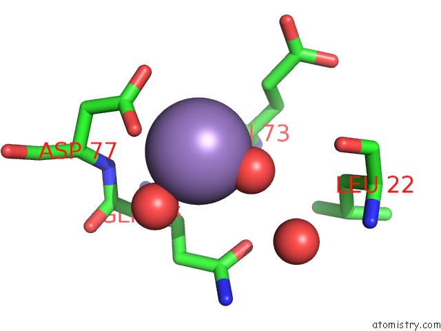
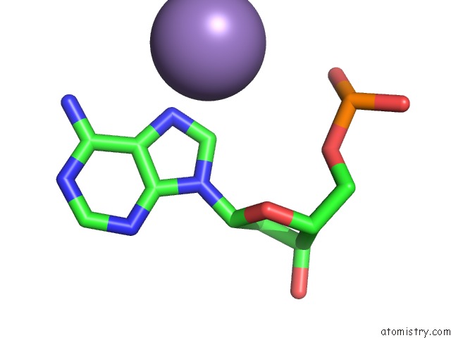
Manganese binding site 1 out of 4 in 5mlu
Go back to
Manganese binding site 1 out
of 4 in the Crystal Structure of the Pfv Gag Cbs Bound to A Mononucleosome
Mono view
Stereo pair view
Mono view
Stereo pair view
A full contact list of Manganese with other atoms in the Mn binding
site number 1 of Crystal Structure of the Pfv Gag Cbs Bound to A Mononucleosome within 5.0Å range:
|
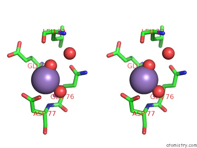
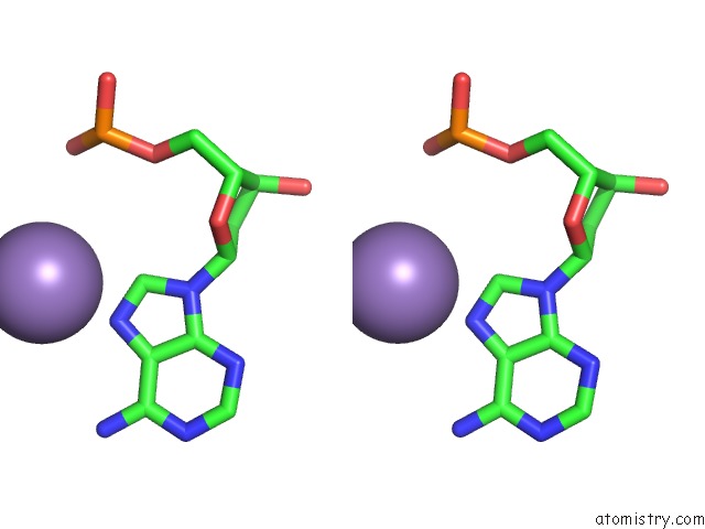
Manganese binding site 2 out of 4 in 5mlu
Go back to
Manganese binding site 2 out
of 4 in the Crystal Structure of the Pfv Gag Cbs Bound to A Mononucleosome
Mono view
Stereo pair view
Mono view
Stereo pair view
A full contact list of Manganese with other atoms in the Mn binding
site number 2 of Crystal Structure of the Pfv Gag Cbs Bound to A Mononucleosome within 5.0Å range:
|
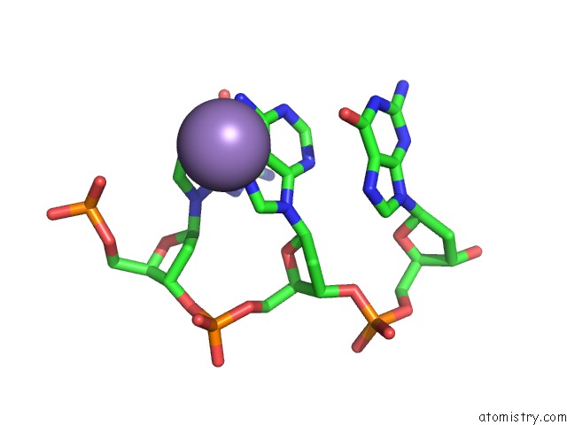
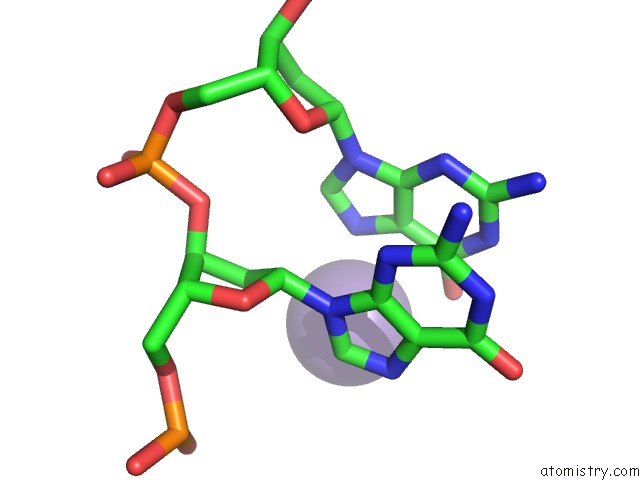
Manganese binding site 3 out of 4 in 5mlu
Go back to
Manganese binding site 3 out
of 4 in the Crystal Structure of the Pfv Gag Cbs Bound to A Mononucleosome
Mono view
Stereo pair view
Mono view
Stereo pair view
A full contact list of Manganese with other atoms in the Mn binding
site number 3 of Crystal Structure of the Pfv Gag Cbs Bound to A Mononucleosome within 5.0Å range:
|
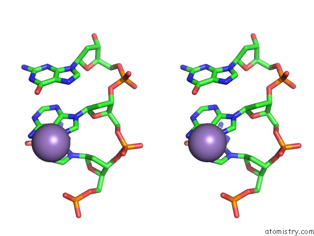
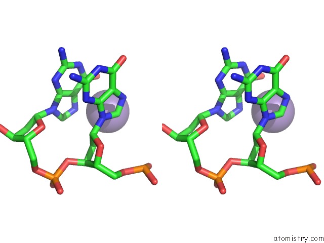
Manganese binding site 4 out of 4 in 5mlu
Go back to
Manganese binding site 4 out
of 4 in the Crystal Structure of the Pfv Gag Cbs Bound to A Mononucleosome
Mono view
Stereo pair view
Mono view
Stereo pair view
A full contact list of Manganese with other atoms in the Mn binding
site number 4 of Crystal Structure of the Pfv Gag Cbs Bound to A Mononucleosome within 5.0Å range:
|
Reference:
P.Lesbats,
E.Serrao,
D.P.Maskell,
V.E.Pye,
N.O'reilly,
D.Lindemann,
A.N.Engelman,
P.Cherepanov.
Structural Basis For Spumavirus Gag Tethering to Chromatin. Proc. Natl. Acad. Sci. V. 114 5509 2017U.S.A..
ISSN: ESSN 1091-6490
PubMed: 28490494
DOI: 10.1073/PNAS.1621159114
Page generated: Sun Oct 6 01:58:36 2024
ISSN: ESSN 1091-6490
PubMed: 28490494
DOI: 10.1073/PNAS.1621159114
Last articles
Zn in 9MJ5Zn in 9HNW
Zn in 9G0L
Zn in 9FNE
Zn in 9DZN
Zn in 9E0I
Zn in 9D32
Zn in 9DAK
Zn in 8ZXC
Zn in 8ZUF