Manganese »
PDB 5lyx-5nh9 »
5mc4 »
Manganese in PDB 5mc4: Crystal Structure of GLY448ARG Mutant of Human Prolidase with Mn Ions and Glypro Ligand
Enzymatic activity of Crystal Structure of GLY448ARG Mutant of Human Prolidase with Mn Ions and Glypro Ligand
All present enzymatic activity of Crystal Structure of GLY448ARG Mutant of Human Prolidase with Mn Ions and Glypro Ligand:
3.4.13.9;
3.4.13.9;
Protein crystallography data
The structure of Crystal Structure of GLY448ARG Mutant of Human Prolidase with Mn Ions and Glypro Ligand, PDB code: 5mc4
was solved by
P.Wilk,
U.Mueller,
H.Dobbek,
M.S.Weiss,
with X-Ray Crystallography technique. A brief refinement statistics is given in the table below:
| Resolution Low / High (Å) | 47.97 / 1.80 |
| Space group | C 2 2 21 |
| Cell size a, b, c (Å), α, β, γ (°) | 103.742, 107.737, 210.878, 90.00, 90.00, 90.00 |
| R / Rfree (%) | 16.3 / 19.2 |
Manganese Binding Sites:
The binding sites of Manganese atom in the Crystal Structure of GLY448ARG Mutant of Human Prolidase with Mn Ions and Glypro Ligand
(pdb code 5mc4). This binding sites where shown within
5.0 Angstroms radius around Manganese atom.
In total 4 binding sites of Manganese where determined in the Crystal Structure of GLY448ARG Mutant of Human Prolidase with Mn Ions and Glypro Ligand, PDB code: 5mc4:
Jump to Manganese binding site number: 1; 2; 3; 4;
In total 4 binding sites of Manganese where determined in the Crystal Structure of GLY448ARG Mutant of Human Prolidase with Mn Ions and Glypro Ligand, PDB code: 5mc4:
Jump to Manganese binding site number: 1; 2; 3; 4;
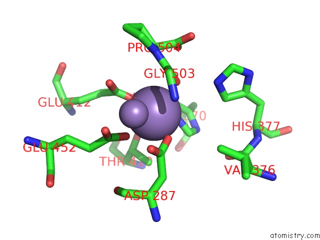
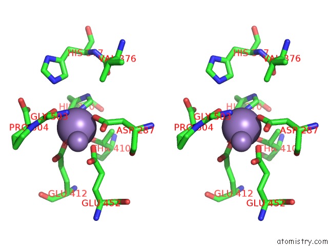
Manganese binding site 1 out of 4 in 5mc4
Go back to
Manganese binding site 1 out
of 4 in the Crystal Structure of GLY448ARG Mutant of Human Prolidase with Mn Ions and Glypro Ligand
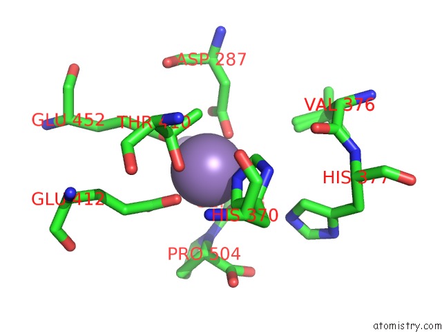
Mono view
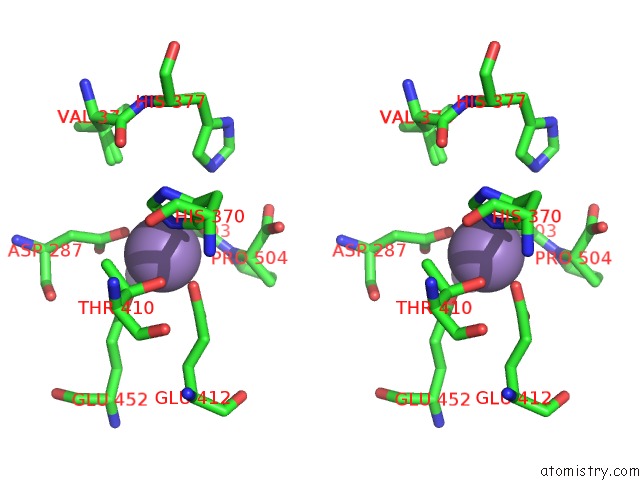
Stereo pair view
Mono view
Stereo pair view
A full contact list of Manganese with other atoms in the Mn binding
site number 1 of Crystal Structure of GLY448ARG Mutant of Human Prolidase with Mn Ions and Glypro Ligand within 5.0Å range:
|
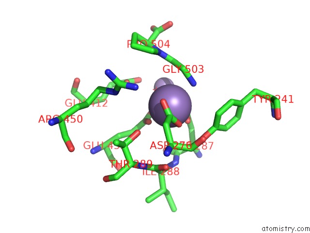
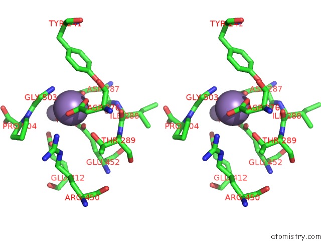
Manganese binding site 2 out of 4 in 5mc4
Go back to
Manganese binding site 2 out
of 4 in the Crystal Structure of GLY448ARG Mutant of Human Prolidase with Mn Ions and Glypro Ligand
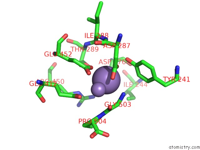
Mono view
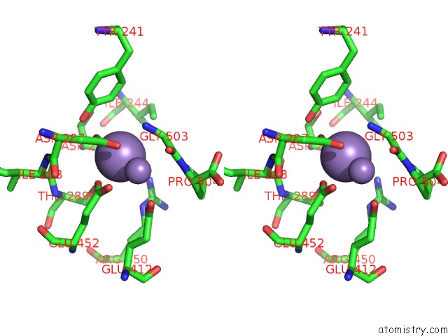
Stereo pair view
Mono view
Stereo pair view
A full contact list of Manganese with other atoms in the Mn binding
site number 2 of Crystal Structure of GLY448ARG Mutant of Human Prolidase with Mn Ions and Glypro Ligand within 5.0Å range:
|
Manganese binding site 3 out of 4 in 5mc4
Go back to
Manganese binding site 3 out
of 4 in the Crystal Structure of GLY448ARG Mutant of Human Prolidase with Mn Ions and Glypro Ligand
Mono view
Stereo pair view
Mono view
Stereo pair view
A full contact list of Manganese with other atoms in the Mn binding
site number 3 of Crystal Structure of GLY448ARG Mutant of Human Prolidase with Mn Ions and Glypro Ligand within 5.0Å range:
|
Manganese binding site 4 out of 4 in 5mc4
Go back to
Manganese binding site 4 out
of 4 in the Crystal Structure of GLY448ARG Mutant of Human Prolidase with Mn Ions and Glypro Ligand
Mono view
Stereo pair view
Mono view
Stereo pair view
A full contact list of Manganese with other atoms in the Mn binding
site number 4 of Crystal Structure of GLY448ARG Mutant of Human Prolidase with Mn Ions and Glypro Ligand within 5.0Å range:
|
Reference:
P.Wilk,
M.Uehlein,
R.Piwowarczyk,
H.Dobbek,
U.Mueller,
M.S.Weiss.
Structural Basis For Prolidase Deficiency Disease Mechanisms. Febs J. V. 285 3422 2018.
ISSN: ISSN 1742-4658
PubMed: 30066404
DOI: 10.1111/FEBS.14620
Page generated: Sun Oct 6 01:57:25 2024
ISSN: ISSN 1742-4658
PubMed: 30066404
DOI: 10.1111/FEBS.14620
Last articles
Zn in 9J0NZn in 9J0O
Zn in 9J0P
Zn in 9FJX
Zn in 9EKB
Zn in 9C0F
Zn in 9CAH
Zn in 9CH0
Zn in 9CH3
Zn in 9CH1