Manganese »
PDB 5hzz-5jpf »
5j8l »
Manganese in PDB 5j8l: Crystal Structure of D-Tagatose 3-Epimerase C66S From Pseudomonas Cichorii in Complex with 1-Deoxy L-Tagatose, Using A Crystal Grown in Microgravity
Enzymatic activity of Crystal Structure of D-Tagatose 3-Epimerase C66S From Pseudomonas Cichorii in Complex with 1-Deoxy L-Tagatose, Using A Crystal Grown in Microgravity
All present enzymatic activity of Crystal Structure of D-Tagatose 3-Epimerase C66S From Pseudomonas Cichorii in Complex with 1-Deoxy L-Tagatose, Using A Crystal Grown in Microgravity:
5.1.3.31;
5.1.3.31;
Protein crystallography data
The structure of Crystal Structure of D-Tagatose 3-Epimerase C66S From Pseudomonas Cichorii in Complex with 1-Deoxy L-Tagatose, Using A Crystal Grown in Microgravity, PDB code: 5j8l
was solved by
H.Yoshida,
A.Yoshihara,
K.Izumori,
S.Kamitori,
with X-Ray Crystallography technique. A brief refinement statistics is given in the table below:
| Resolution Low / High (Å) | 38.68 / 1.73 |
| Space group | P 1 21 1 |
| Cell size a, b, c (Å), α, β, γ (°) | 52.580, 126.535, 98.903, 90.00, 101.45, 90.00 |
| R / Rfree (%) | 19.4 / 21.9 |
Manganese Binding Sites:
The binding sites of Manganese atom in the Crystal Structure of D-Tagatose 3-Epimerase C66S From Pseudomonas Cichorii in Complex with 1-Deoxy L-Tagatose, Using A Crystal Grown in Microgravity
(pdb code 5j8l). This binding sites where shown within
5.0 Angstroms radius around Manganese atom.
In total 4 binding sites of Manganese where determined in the Crystal Structure of D-Tagatose 3-Epimerase C66S From Pseudomonas Cichorii in Complex with 1-Deoxy L-Tagatose, Using A Crystal Grown in Microgravity, PDB code: 5j8l:
Jump to Manganese binding site number: 1; 2; 3; 4;
In total 4 binding sites of Manganese where determined in the Crystal Structure of D-Tagatose 3-Epimerase C66S From Pseudomonas Cichorii in Complex with 1-Deoxy L-Tagatose, Using A Crystal Grown in Microgravity, PDB code: 5j8l:
Jump to Manganese binding site number: 1; 2; 3; 4;
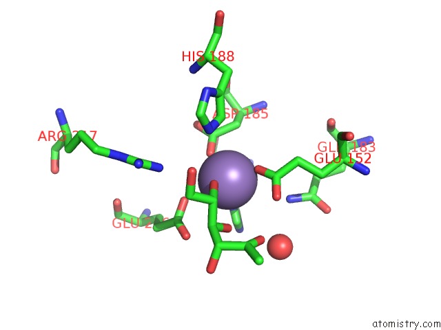
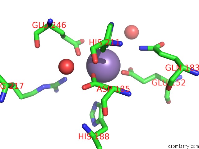
Manganese binding site 1 out of 4 in 5j8l
Go back to
Manganese binding site 1 out
of 4 in the Crystal Structure of D-Tagatose 3-Epimerase C66S From Pseudomonas Cichorii in Complex with 1-Deoxy L-Tagatose, Using A Crystal Grown in Microgravity
Mono view
Stereo pair view
Mono view
Stereo pair view
A full contact list of Manganese with other atoms in the Mn binding
site number 1 of Crystal Structure of D-Tagatose 3-Epimerase C66S From Pseudomonas Cichorii in Complex with 1-Deoxy L-Tagatose, Using A Crystal Grown in Microgravity within 5.0Å range:
|
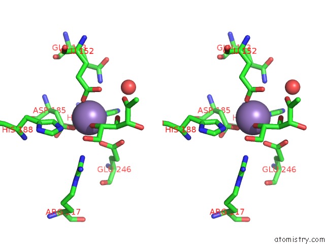
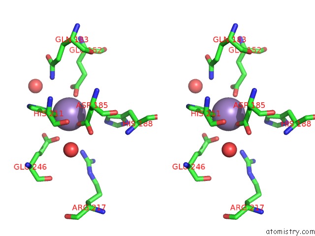
Manganese binding site 2 out of 4 in 5j8l
Go back to
Manganese binding site 2 out
of 4 in the Crystal Structure of D-Tagatose 3-Epimerase C66S From Pseudomonas Cichorii in Complex with 1-Deoxy L-Tagatose, Using A Crystal Grown in Microgravity
Mono view
Stereo pair view
Mono view
Stereo pair view
A full contact list of Manganese with other atoms in the Mn binding
site number 2 of Crystal Structure of D-Tagatose 3-Epimerase C66S From Pseudomonas Cichorii in Complex with 1-Deoxy L-Tagatose, Using A Crystal Grown in Microgravity within 5.0Å range:
|
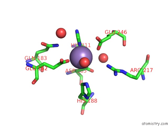
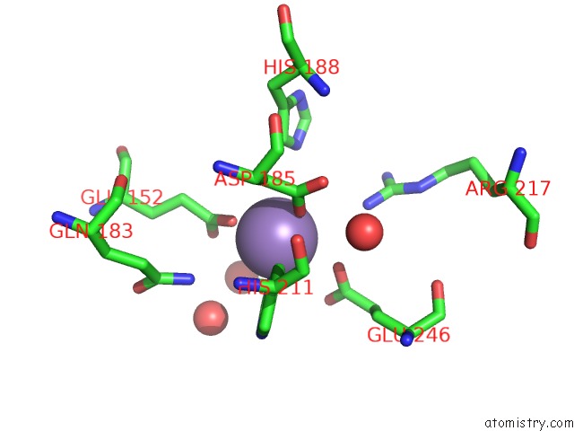
Manganese binding site 3 out of 4 in 5j8l
Go back to
Manganese binding site 3 out
of 4 in the Crystal Structure of D-Tagatose 3-Epimerase C66S From Pseudomonas Cichorii in Complex with 1-Deoxy L-Tagatose, Using A Crystal Grown in Microgravity
Mono view
Stereo pair view
Mono view
Stereo pair view
A full contact list of Manganese with other atoms in the Mn binding
site number 3 of Crystal Structure of D-Tagatose 3-Epimerase C66S From Pseudomonas Cichorii in Complex with 1-Deoxy L-Tagatose, Using A Crystal Grown in Microgravity within 5.0Å range:
|
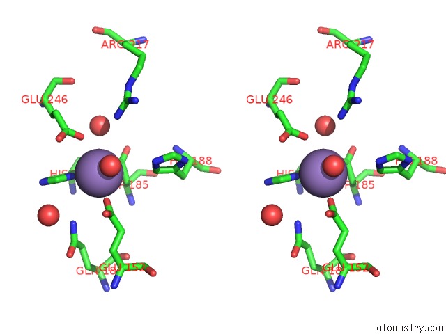
Manganese binding site 4 out of 4 in 5j8l
Go back to
Manganese binding site 4 out
of 4 in the Crystal Structure of D-Tagatose 3-Epimerase C66S From Pseudomonas Cichorii in Complex with 1-Deoxy L-Tagatose, Using A Crystal Grown in Microgravity
Mono view
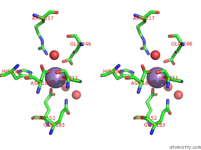
Stereo pair view
Mono view
Stereo pair view
A full contact list of Manganese with other atoms in the Mn binding
site number 4 of Crystal Structure of D-Tagatose 3-Epimerase C66S From Pseudomonas Cichorii in Complex with 1-Deoxy L-Tagatose, Using A Crystal Grown in Microgravity within 5.0Å range:
|
Reference:
H.Yoshida,
A.Yoshihara,
T.Ishii,
K.Izumori,
S.Kamitori.
X-Ray Structures of the Pseudomonas Cichorii D-Tagatose 3-Epimerase Mutant Form C66S Recognizing Deoxy Sugars As Substrates Appl. Microbiol. Biotechnol. V. 100 10403 2016.
ISSN: ESSN 1432-0614
PubMed: 27368739
DOI: 10.1007/S00253-016-7673-7
Page generated: Sun Oct 6 01:29:47 2024
ISSN: ESSN 1432-0614
PubMed: 27368739
DOI: 10.1007/S00253-016-7673-7
Last articles
Zn in 9JYWZn in 9IR4
Zn in 9IR3
Zn in 9GMX
Zn in 9GMW
Zn in 9JEJ
Zn in 9ERF
Zn in 9ERE
Zn in 9EGV
Zn in 9EGW