Manganese »
PDB 5hzz-5jpf »
5iw0 »
Manganese in PDB 5iw0: Linked KDM5A Jmj Domain Bound to the Inhibitor N19 [2-(5-((4-Chloro-2- Methylbenzyl)Oxy)-1H-Pyrazol-1-Yl)Isonicotinic Acid]
Protein crystallography data
The structure of Linked KDM5A Jmj Domain Bound to the Inhibitor N19 [2-(5-((4-Chloro-2- Methylbenzyl)Oxy)-1H-Pyrazol-1-Yl)Isonicotinic Acid], PDB code: 5iw0
was solved by
J.R.Horton,
X.Cheng,
with X-Ray Crystallography technique. A brief refinement statistics is given in the table below:
| Resolution Low / High (Å) | 32.97 / 1.63 |
| Space group | C 1 2 1 |
| Cell size a, b, c (Å), α, β, γ (°) | 116.320, 62.741, 46.609, 90.00, 91.93, 90.00 |
| R / Rfree (%) | 15.7 / 19.2 |
Other elements in 5iw0:
The structure of Linked KDM5A Jmj Domain Bound to the Inhibitor N19 [2-(5-((4-Chloro-2- Methylbenzyl)Oxy)-1H-Pyrazol-1-Yl)Isonicotinic Acid] also contains other interesting chemical elements:
| Chlorine | (Cl) | 1 atom |
Manganese Binding Sites:
The binding sites of Manganese atom in the Linked KDM5A Jmj Domain Bound to the Inhibitor N19 [2-(5-((4-Chloro-2- Methylbenzyl)Oxy)-1H-Pyrazol-1-Yl)Isonicotinic Acid]
(pdb code 5iw0). This binding sites where shown within
5.0 Angstroms radius around Manganese atom.
In total only one binding site of Manganese was determined in the Linked KDM5A Jmj Domain Bound to the Inhibitor N19 [2-(5-((4-Chloro-2- Methylbenzyl)Oxy)-1H-Pyrazol-1-Yl)Isonicotinic Acid], PDB code: 5iw0:
In total only one binding site of Manganese was determined in the Linked KDM5A Jmj Domain Bound to the Inhibitor N19 [2-(5-((4-Chloro-2- Methylbenzyl)Oxy)-1H-Pyrazol-1-Yl)Isonicotinic Acid], PDB code: 5iw0:
Manganese binding site 1 out of 1 in 5iw0
Go back to
Manganese binding site 1 out
of 1 in the Linked KDM5A Jmj Domain Bound to the Inhibitor N19 [2-(5-((4-Chloro-2- Methylbenzyl)Oxy)-1H-Pyrazol-1-Yl)Isonicotinic Acid]
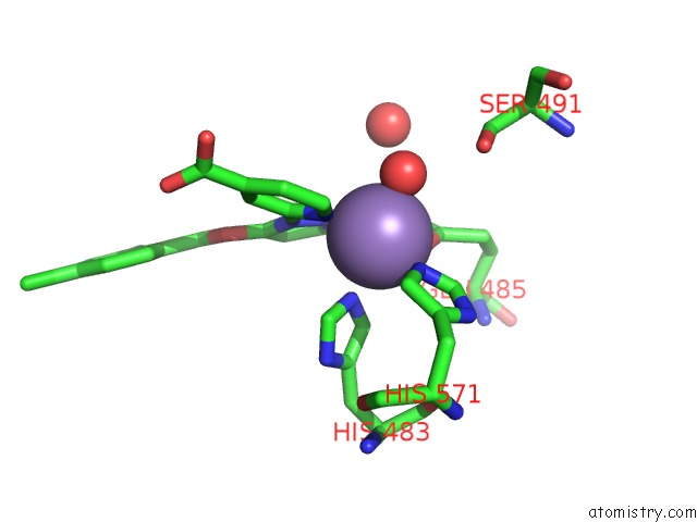
Mono view
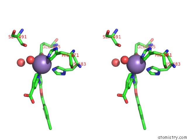
Stereo pair view
Mono view
Stereo pair view
A full contact list of Manganese with other atoms in the Mn binding
site number 1 of Linked KDM5A Jmj Domain Bound to the Inhibitor N19 [2-(5-((4-Chloro-2- Methylbenzyl)Oxy)-1H-Pyrazol-1-Yl)Isonicotinic Acid] within 5.0Å range:
|
Reference:
J.R.Horton,
X.Liu,
M.Gale,
L.Wu,
J.R.Shanks,
X.Zhang,
P.J.Webber,
J.S.Bell,
S.C.Kales,
B.T.Mott,
G.Rai,
D.J.Jansen,
M.J.Henderson,
D.J.Urban,
M.D.Hall,
A.Simeonov,
D.J.Maloney,
M.A.Johns,
H.Fu,
A.Jadhav,
P.M.Vertino,
Q.Yan,
X.Cheng.
Structural Basis For KDM5A Histone Lysine Demethylase Inhibition By Diverse Compounds. Cell Chem Biol V. 23 769 2016.
ISSN: ESSN 2451-9456
PubMed: 27427228
DOI: 10.1016/J.CHEMBIOL.2016.06.006
Page generated: Sun Oct 6 01:28:17 2024
ISSN: ESSN 2451-9456
PubMed: 27427228
DOI: 10.1016/J.CHEMBIOL.2016.06.006
Last articles
Zn in 9JYWZn in 9IR4
Zn in 9IR3
Zn in 9GMX
Zn in 9GMW
Zn in 9JEJ
Zn in 9ERF
Zn in 9ERE
Zn in 9EGV
Zn in 9EGW