Manganese »
PDB 5gjc-5hwu »
5hn4 »
Manganese in PDB 5hn4: Crystal Structure of Beta-Decarboxylating Dehydrogenase (TK0280) From Thermococcus Kodakarensis Complexed with Mn and Homoisocitrate
Protein crystallography data
The structure of Crystal Structure of Beta-Decarboxylating Dehydrogenase (TK0280) From Thermococcus Kodakarensis Complexed with Mn and Homoisocitrate, PDB code: 5hn4
was solved by
T.Shimizu,
T.Tomita,
M.Nishiyama,
with X-Ray Crystallography technique. A brief refinement statistics is given in the table below:
| Resolution Low / High (Å) | 143.23 / 2.64 |
| Space group | P 43 21 2 |
| Cell size a, b, c (Å), α, β, γ (°) | 113.669, 113.669, 143.226, 90.00, 90.00, 90.00 |
| R / Rfree (%) | 20.1 / 22 |
Manganese Binding Sites:
The binding sites of Manganese atom in the Crystal Structure of Beta-Decarboxylating Dehydrogenase (TK0280) From Thermococcus Kodakarensis Complexed with Mn and Homoisocitrate
(pdb code 5hn4). This binding sites where shown within
5.0 Angstroms radius around Manganese atom.
In total only one binding site of Manganese was determined in the Crystal Structure of Beta-Decarboxylating Dehydrogenase (TK0280) From Thermococcus Kodakarensis Complexed with Mn and Homoisocitrate, PDB code: 5hn4:
In total only one binding site of Manganese was determined in the Crystal Structure of Beta-Decarboxylating Dehydrogenase (TK0280) From Thermococcus Kodakarensis Complexed with Mn and Homoisocitrate, PDB code: 5hn4:
Manganese binding site 1 out of 1 in 5hn4
Go back to
Manganese binding site 1 out
of 1 in the Crystal Structure of Beta-Decarboxylating Dehydrogenase (TK0280) From Thermococcus Kodakarensis Complexed with Mn and Homoisocitrate
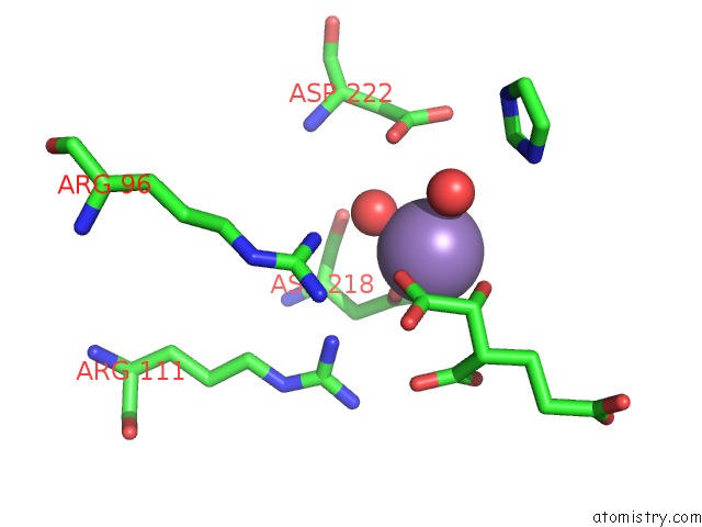
Mono view
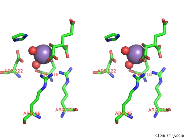
Stereo pair view
Mono view
Stereo pair view
A full contact list of Manganese with other atoms in the Mn binding
site number 1 of Crystal Structure of Beta-Decarboxylating Dehydrogenase (TK0280) From Thermococcus Kodakarensis Complexed with Mn and Homoisocitrate within 5.0Å range:
|
Reference:
T.Shimizu,
L.Yin,
A.Yoshida,
Y.Yokooji,
S.I.Hachisuka,
T.Sato,
T.Tomita,
H.Nishida,
H.Atomi,
T.Kuzuyama,
M.Nishiyama.
Structure and Function of An Ancestral-Type Beta-Decarboxylating Dehydrogenase From Thermococcus Kodakarensis Biochem. J. V. 474 105 2017.
ISSN: ESSN 1470-8728
PubMed: 27831491
DOI: 10.1042/BCJ20160699
Page generated: Sun Oct 6 00:30:50 2024
ISSN: ESSN 1470-8728
PubMed: 27831491
DOI: 10.1042/BCJ20160699
Last articles
Zn in 9J0NZn in 9J0O
Zn in 9J0P
Zn in 9FJX
Zn in 9EKB
Zn in 9C0F
Zn in 9CAH
Zn in 9CH0
Zn in 9CH3
Zn in 9CH1