Manganese »
PDB 5gjc-5hwu »
5h0o »
Manganese in PDB 5h0o: Crystal Structure of Deep-Sea Thermophilic Bacteriophage GVE2 Hnh Endonuclease with Manganese Ion
Protein crystallography data
The structure of Crystal Structure of Deep-Sea Thermophilic Bacteriophage GVE2 Hnh Endonuclease with Manganese Ion, PDB code: 5h0o
was solved by
L.K.Zhang,
D.D.Xu,
Y.C.Huang,
Y.Gong,
with X-Ray Crystallography technique. A brief refinement statistics is given in the table below:
| Resolution Low / High (Å) | 25.86 / 1.53 |
| Space group | P 43 21 2 |
| Cell size a, b, c (Å), α, β, γ (°) | 66.847, 66.847, 51.511, 90.00, 90.00, 90.00 |
| R / Rfree (%) | 14.1 / 17.4 |
Manganese Binding Sites:
The binding sites of Manganese atom in the Crystal Structure of Deep-Sea Thermophilic Bacteriophage GVE2 Hnh Endonuclease with Manganese Ion
(pdb code 5h0o). This binding sites where shown within
5.0 Angstroms radius around Manganese atom.
In total only one binding site of Manganese was determined in the Crystal Structure of Deep-Sea Thermophilic Bacteriophage GVE2 Hnh Endonuclease with Manganese Ion, PDB code: 5h0o:
In total only one binding site of Manganese was determined in the Crystal Structure of Deep-Sea Thermophilic Bacteriophage GVE2 Hnh Endonuclease with Manganese Ion, PDB code: 5h0o:
Manganese binding site 1 out of 1 in 5h0o
Go back to
Manganese binding site 1 out
of 1 in the Crystal Structure of Deep-Sea Thermophilic Bacteriophage GVE2 Hnh Endonuclease with Manganese Ion
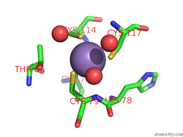
Mono view
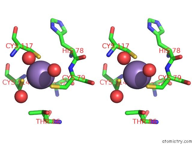
Stereo pair view
Mono view
Stereo pair view
A full contact list of Manganese with other atoms in the Mn binding
site number 1 of Crystal Structure of Deep-Sea Thermophilic Bacteriophage GVE2 Hnh Endonuclease with Manganese Ion within 5.0Å range:
|
Reference:
L.K.Zhang,
D.D.Xu,
Y.C.Huang,
X.Y.Zhu,
M.W.Rui,
T.Wan,
X.Zheng,
Y.L.Shen,
X.D.Chen,
K.S.Ma,
Y.Gong.
Structural and Functional Characterization of Deep-Sea Thermophilic Bacteriophage GVE2 Hnh Endonuclease. Sci Rep V. 7 42542 2017.
ISSN: ESSN 2045-2322
PubMed: 28211904
DOI: 10.1038/SREP42542
Page generated: Sat Aug 16 17:35:07 2025
ISSN: ESSN 2045-2322
PubMed: 28211904
DOI: 10.1038/SREP42542
Last articles
Mo in 3K7RMo in 3ML1
Mo in 3K6X
Mo in 3L4P
Mo in 3K1A
Mo in 3K6W
Mo in 3GZG
Mo in 3HRD
Mo in 3IR5
Mo in 3IR7