Manganese »
PDB 5ekw-5fxv »
5fpv »
Manganese in PDB 5fpv: Crystal Structure of Human JMJD2A in Complex with Compound KDOAM20A
Enzymatic activity of Crystal Structure of Human JMJD2A in Complex with Compound KDOAM20A
All present enzymatic activity of Crystal Structure of Human JMJD2A in Complex with Compound KDOAM20A:
1.14.11.27;
1.14.11.27;
Protein crystallography data
The structure of Crystal Structure of Human JMJD2A in Complex with Compound KDOAM20A, PDB code: 5fpv
was solved by
V.Srikannathasan,
C.Gileadi,
F.Von Delft,
C.H.Arrowsmith,
C.Bountra,
A.Edwards,
U.Oppermann,
with X-Ray Crystallography technique. A brief refinement statistics is given in the table below:
| Resolution Low / High (Å) | 85.327 / 2.44 |
| Space group | P 1 21 1 |
| Cell size a, b, c (Å), α, β, γ (°) | 111.346, 103.509, 157.103, 90.00, 106.37, 90.00 |
| R / Rfree (%) | 19.55 / 22.89 |
Other elements in 5fpv:
The structure of Crystal Structure of Human JMJD2A in Complex with Compound KDOAM20A also contains other interesting chemical elements:
| Nickel | (Ni) | 15 atoms |
| Zinc | (Zn) | 8 atoms |
Manganese Binding Sites:
The binding sites of Manganese atom in the Crystal Structure of Human JMJD2A in Complex with Compound KDOAM20A
(pdb code 5fpv). This binding sites where shown within
5.0 Angstroms radius around Manganese atom.
In total 8 binding sites of Manganese where determined in the Crystal Structure of Human JMJD2A in Complex with Compound KDOAM20A, PDB code: 5fpv:
Jump to Manganese binding site number: 1; 2; 3; 4; 5; 6; 7; 8;
In total 8 binding sites of Manganese where determined in the Crystal Structure of Human JMJD2A in Complex with Compound KDOAM20A, PDB code: 5fpv:
Jump to Manganese binding site number: 1; 2; 3; 4; 5; 6; 7; 8;
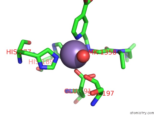
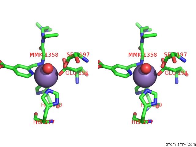
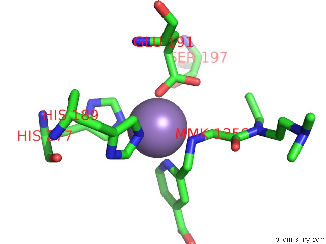
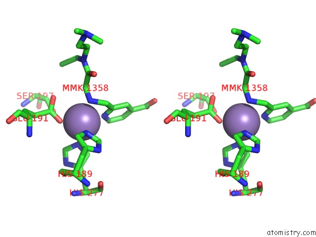
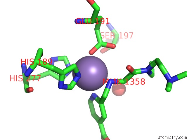
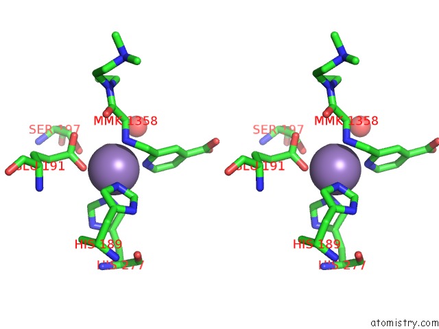
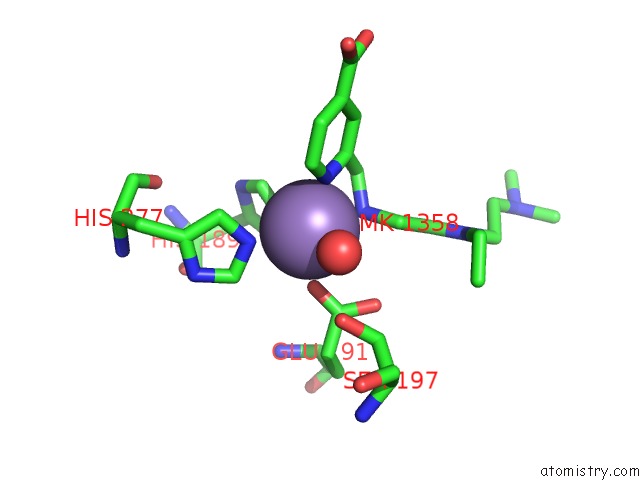
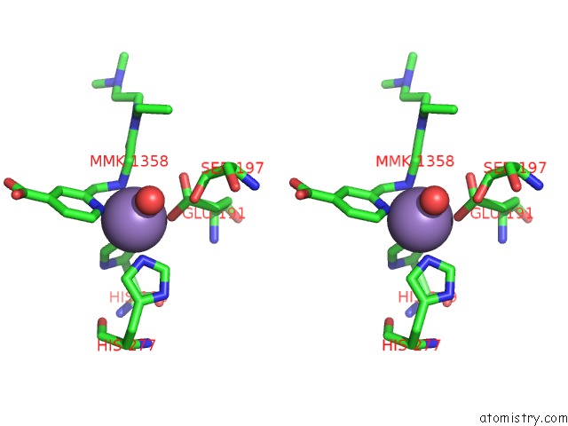
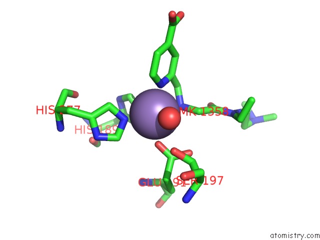
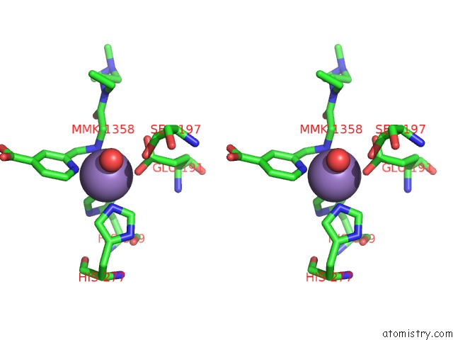
Manganese binding site 1 out of 8 in 5fpv
Go back to
Manganese binding site 1 out
of 8 in the Crystal Structure of Human JMJD2A in Complex with Compound KDOAM20A
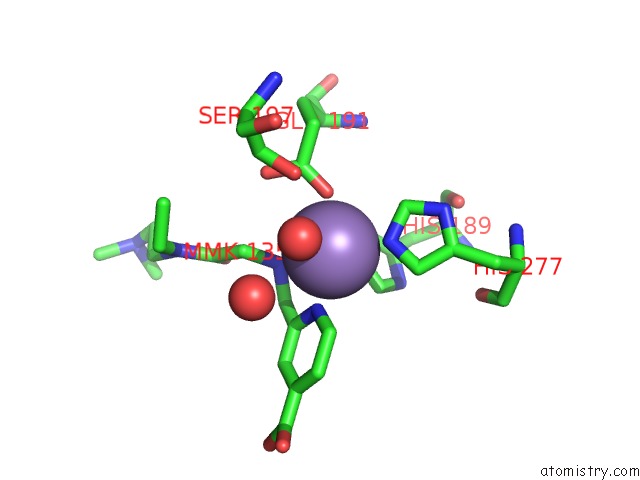
Mono view
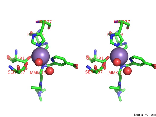
Stereo pair view
Mono view
Stereo pair view
A full contact list of Manganese with other atoms in the Mn binding
site number 1 of Crystal Structure of Human JMJD2A in Complex with Compound KDOAM20A within 5.0Å range:
|
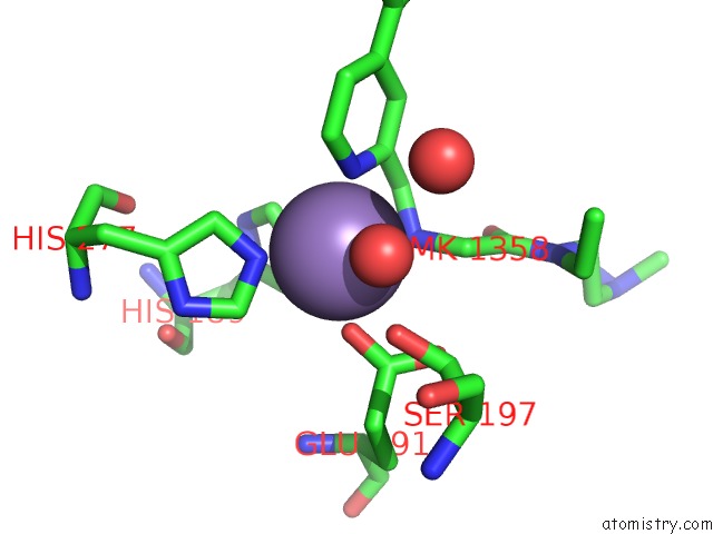
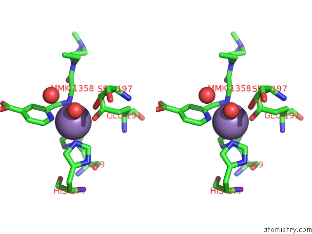
Manganese binding site 2 out of 8 in 5fpv
Go back to
Manganese binding site 2 out
of 8 in the Crystal Structure of Human JMJD2A in Complex with Compound KDOAM20A
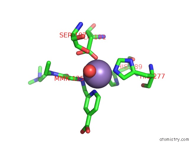
Mono view
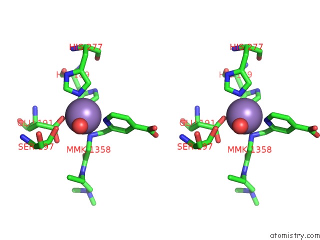
Stereo pair view
Mono view
Stereo pair view
A full contact list of Manganese with other atoms in the Mn binding
site number 2 of Crystal Structure of Human JMJD2A in Complex with Compound KDOAM20A within 5.0Å range:
|
Manganese binding site 3 out of 8 in 5fpv
Go back to
Manganese binding site 3 out
of 8 in the Crystal Structure of Human JMJD2A in Complex with Compound KDOAM20A
Mono view
Stereo pair view
Mono view
Stereo pair view
A full contact list of Manganese with other atoms in the Mn binding
site number 3 of Crystal Structure of Human JMJD2A in Complex with Compound KDOAM20A within 5.0Å range:
|
Manganese binding site 4 out of 8 in 5fpv
Go back to
Manganese binding site 4 out
of 8 in the Crystal Structure of Human JMJD2A in Complex with Compound KDOAM20A
Mono view
Stereo pair view
Mono view
Stereo pair view
A full contact list of Manganese with other atoms in the Mn binding
site number 4 of Crystal Structure of Human JMJD2A in Complex with Compound KDOAM20A within 5.0Å range:
|
Manganese binding site 5 out of 8 in 5fpv
Go back to
Manganese binding site 5 out
of 8 in the Crystal Structure of Human JMJD2A in Complex with Compound KDOAM20A
Mono view
Stereo pair view
Mono view
Stereo pair view
A full contact list of Manganese with other atoms in the Mn binding
site number 5 of Crystal Structure of Human JMJD2A in Complex with Compound KDOAM20A within 5.0Å range:
|
Manganese binding site 6 out of 8 in 5fpv
Go back to
Manganese binding site 6 out
of 8 in the Crystal Structure of Human JMJD2A in Complex with Compound KDOAM20A
Mono view
Stereo pair view
Mono view
Stereo pair view
A full contact list of Manganese with other atoms in the Mn binding
site number 6 of Crystal Structure of Human JMJD2A in Complex with Compound KDOAM20A within 5.0Å range:
|
Manganese binding site 7 out of 8 in 5fpv
Go back to
Manganese binding site 7 out
of 8 in the Crystal Structure of Human JMJD2A in Complex with Compound KDOAM20A
Mono view
Stereo pair view
Mono view
Stereo pair view
A full contact list of Manganese with other atoms in the Mn binding
site number 7 of Crystal Structure of Human JMJD2A in Complex with Compound KDOAM20A within 5.0Å range:
|
Manganese binding site 8 out of 8 in 5fpv
Go back to
Manganese binding site 8 out
of 8 in the Crystal Structure of Human JMJD2A in Complex with Compound KDOAM20A
Mono view
Stereo pair view
Mono view
Stereo pair view
A full contact list of Manganese with other atoms in the Mn binding
site number 8 of Crystal Structure of Human JMJD2A in Complex with Compound KDOAM20A within 5.0Å range:
|
Reference:
C.Johansson,
S.Velupillai,
A.Tumber,
A.Szykowska,
E.S.Hookway,
R.P.Nowak,
C.Strain-Damerell,
C.Gileadi,
M.Philpott,
N.Burgess-Brown,
N.Wu,
J.Kopec,
A.Nuzzi,
H.Steuber,
U.Egner,
V.Badock,
S.Munro,
N.B.Lathangue,
S.Westaway,
J.Brown,
N.Athanasou,
R.Prinjha,
P.E.Brennan,
U.Oppermann.
Structural Analysis of Human KDM5B Guides Histone Demethylase Inhibitor Development. Nat.Chem.Biol. V. 12 539 2016.
ISSN: ISSN 1552-4450
PubMed: 27214403
DOI: 10.1038/NCHEMBIO.2087
Page generated: Sun Oct 6 00:15:14 2024
ISSN: ISSN 1552-4450
PubMed: 27214403
DOI: 10.1038/NCHEMBIO.2087
Last articles
Zn in 9J0NZn in 9J0O
Zn in 9J0P
Zn in 9FJX
Zn in 9EKB
Zn in 9C0F
Zn in 9CAH
Zn in 9CH0
Zn in 9CH3
Zn in 9CH1