Manganese »
PDB 5ekw-5fxv »
5fdd »
Manganese in PDB 5fdd: Endonuclease Inhibitor 1 Bound to Influenza Strain H1N1 Polymerase Acidic Subunit N-Terminal Region at pH 7.0
Protein crystallography data
The structure of Endonuclease Inhibitor 1 Bound to Influenza Strain H1N1 Polymerase Acidic Subunit N-Terminal Region at pH 7.0, PDB code: 5fdd
was solved by
S.Fudo,
N.Yamamoto,
M.Nukaga,
T.Odagiri,
M.Tashiro,
T.Hoshino,
with X-Ray Crystallography technique. A brief refinement statistics is given in the table below:
| Resolution Low / High (Å) | 37.79 / 2.51 |
| Space group | P 41 21 2 |
| Cell size a, b, c (Å), α, β, γ (°) | 66.397, 66.397, 127.392, 90.00, 90.00, 90.00 |
| R / Rfree (%) | 20.1 / 23.2 |
Other elements in 5fdd:
The structure of Endonuclease Inhibitor 1 Bound to Influenza Strain H1N1 Polymerase Acidic Subunit N-Terminal Region at pH 7.0 also contains other interesting chemical elements:
| Chlorine | (Cl) | 1 atom |
Manganese Binding Sites:
The binding sites of Manganese atom in the Endonuclease Inhibitor 1 Bound to Influenza Strain H1N1 Polymerase Acidic Subunit N-Terminal Region at pH 7.0
(pdb code 5fdd). This binding sites where shown within
5.0 Angstroms radius around Manganese atom.
In total only one binding site of Manganese was determined in the Endonuclease Inhibitor 1 Bound to Influenza Strain H1N1 Polymerase Acidic Subunit N-Terminal Region at pH 7.0, PDB code: 5fdd:
In total only one binding site of Manganese was determined in the Endonuclease Inhibitor 1 Bound to Influenza Strain H1N1 Polymerase Acidic Subunit N-Terminal Region at pH 7.0, PDB code: 5fdd:
Manganese binding site 1 out of 1 in 5fdd
Go back to
Manganese binding site 1 out
of 1 in the Endonuclease Inhibitor 1 Bound to Influenza Strain H1N1 Polymerase Acidic Subunit N-Terminal Region at pH 7.0
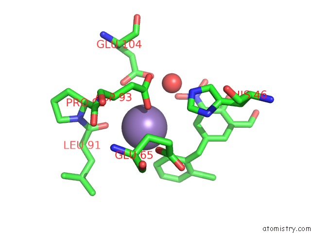
Mono view
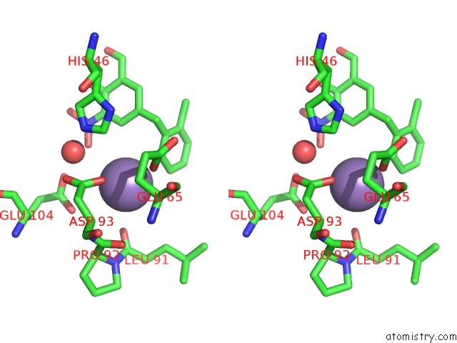
Stereo pair view
Mono view
Stereo pair view
A full contact list of Manganese with other atoms in the Mn binding
site number 1 of Endonuclease Inhibitor 1 Bound to Influenza Strain H1N1 Polymerase Acidic Subunit N-Terminal Region at pH 7.0 within 5.0Å range:
|
Reference:
S.Fudo,
N.Yamamoto,
M.Nukaga,
T.Odagiri,
M.Tashiro,
T.Hoshino.
Two Distinctive Binding Modes of Endonuclease Inhibitors to the N-Terminal Region of Influenza Virus Polymerase Acidic Subunit Biochemistry V. 55 2646 2016.
ISSN: ISSN 0006-2960
PubMed: 27088785
DOI: 10.1021/ACS.BIOCHEM.5B01087
Page generated: Sun Oct 6 00:12:09 2024
ISSN: ISSN 0006-2960
PubMed: 27088785
DOI: 10.1021/ACS.BIOCHEM.5B01087
Last articles
Zn in 9MJ5Zn in 9HNW
Zn in 9G0L
Zn in 9FNE
Zn in 9DZN
Zn in 9E0I
Zn in 9D32
Zn in 9DAK
Zn in 8ZXC
Zn in 8ZUF