Manganese »
PDB 5ekw-5fxv »
5f56 »
Manganese in PDB 5f56: Structure of Recj Complexed with Dna and Ssb-Ct
Protein crystallography data
The structure of Structure of Recj Complexed with Dna and Ssb-Ct, PDB code: 5f56
was solved by
Y.Zhao,
Y.Hua,
K.Cheng,
with X-Ray Crystallography technique. A brief refinement statistics is given in the table below:
| Resolution Low / High (Å) | 29.05 / 2.30 |
| Space group | P 32 2 1 |
| Cell size a, b, c (Å), α, β, γ (°) | 102.220, 102.220, 166.120, 90.00, 90.00, 120.00 |
| R / Rfree (%) | 22.6 / 23.9 |
Manganese Binding Sites:
The binding sites of Manganese atom in the Structure of Recj Complexed with Dna and Ssb-Ct
(pdb code 5f56). This binding sites where shown within
5.0 Angstroms radius around Manganese atom.
In total 2 binding sites of Manganese where determined in the Structure of Recj Complexed with Dna and Ssb-Ct, PDB code: 5f56:
Jump to Manganese binding site number: 1; 2;
In total 2 binding sites of Manganese where determined in the Structure of Recj Complexed with Dna and Ssb-Ct, PDB code: 5f56:
Jump to Manganese binding site number: 1; 2;
Manganese binding site 1 out of 2 in 5f56
Go back to
Manganese binding site 1 out
of 2 in the Structure of Recj Complexed with Dna and Ssb-Ct
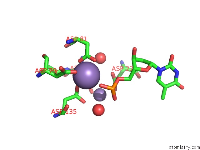
Mono view
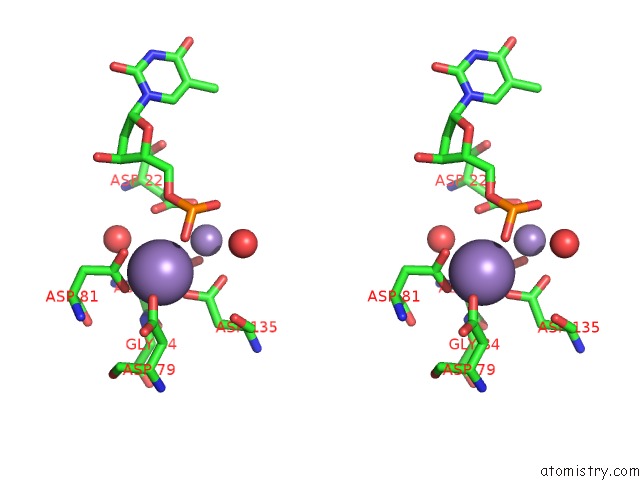
Stereo pair view
Mono view
Stereo pair view
A full contact list of Manganese with other atoms in the Mn binding
site number 1 of Structure of Recj Complexed with Dna and Ssb-Ct within 5.0Å range:
|
Manganese binding site 2 out of 2 in 5f56
Go back to
Manganese binding site 2 out
of 2 in the Structure of Recj Complexed with Dna and Ssb-Ct
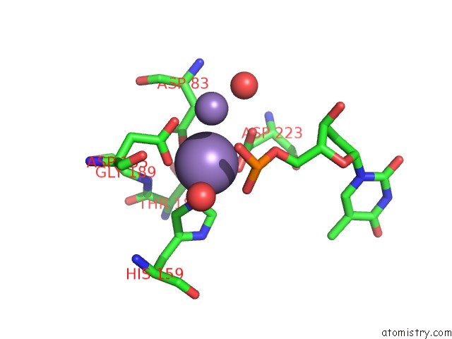
Mono view
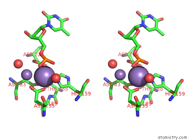
Stereo pair view
Mono view
Stereo pair view
A full contact list of Manganese with other atoms in the Mn binding
site number 2 of Structure of Recj Complexed with Dna and Ssb-Ct within 5.0Å range:
|
Reference:
K.Cheng,
H.Xu,
X.Chen,
L.Wang,
B.Tian,
Y.Zhao,
Y.Hua.
Structural Basis For Dna 5 -End Resection By Recj Elife V. 5 14294 2016.
ISSN: ESSN 2050-084X
PubMed: 27058167
DOI: 10.7554/ELIFE.14294
Page generated: Sun Oct 6 00:11:20 2024
ISSN: ESSN 2050-084X
PubMed: 27058167
DOI: 10.7554/ELIFE.14294
Last articles
Zn in 9MJ5Zn in 9HNW
Zn in 9G0L
Zn in 9FNE
Zn in 9DZN
Zn in 9E0I
Zn in 9D32
Zn in 9DAK
Zn in 8ZXC
Zn in 8ZUF