Manganese »
PDB 5ekw-5fxv »
5f1m »
Manganese in PDB 5f1m: Crystal Structure of Serine/Threonine Phosphatase STP1 From Staphylococcus Aureus
Enzymatic activity of Crystal Structure of Serine/Threonine Phosphatase STP1 From Staphylococcus Aureus
All present enzymatic activity of Crystal Structure of Serine/Threonine Phosphatase STP1 From Staphylococcus Aureus:
3.1.3.16;
3.1.3.16;
Protein crystallography data
The structure of Crystal Structure of Serine/Threonine Phosphatase STP1 From Staphylococcus Aureus, PDB code: 5f1m
was solved by
W.H.Zheng,
T.Wang,
Z.G.Li,
with X-Ray Crystallography technique. A brief refinement statistics is given in the table below:
| Resolution Low / High (Å) | 32.11 / 2.32 |
| Space group | P 2 21 21 |
| Cell size a, b, c (Å), α, β, γ (°) | 38.770, 77.350, 85.360, 90.00, 90.00, 90.00 |
| R / Rfree (%) | 20.2 / 25.6 |
Manganese Binding Sites:
The binding sites of Manganese atom in the Crystal Structure of Serine/Threonine Phosphatase STP1 From Staphylococcus Aureus
(pdb code 5f1m). This binding sites where shown within
5.0 Angstroms radius around Manganese atom.
In total 4 binding sites of Manganese where determined in the Crystal Structure of Serine/Threonine Phosphatase STP1 From Staphylococcus Aureus, PDB code: 5f1m:
Jump to Manganese binding site number: 1; 2; 3; 4;
In total 4 binding sites of Manganese where determined in the Crystal Structure of Serine/Threonine Phosphatase STP1 From Staphylococcus Aureus, PDB code: 5f1m:
Jump to Manganese binding site number: 1; 2; 3; 4;
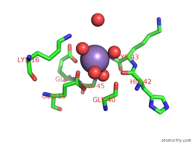
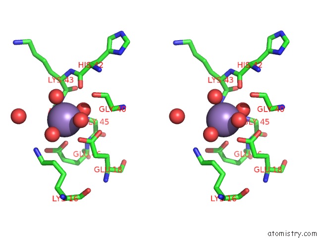
Manganese binding site 1 out of 4 in 5f1m
Go back to
Manganese binding site 1 out
of 4 in the Crystal Structure of Serine/Threonine Phosphatase STP1 From Staphylococcus Aureus
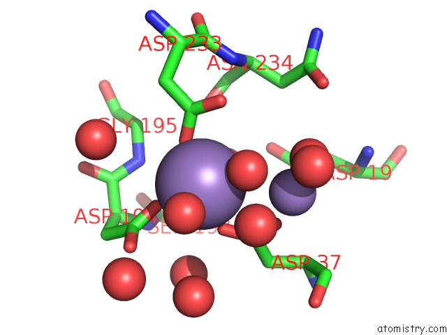
Mono view
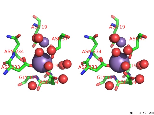
Stereo pair view
Mono view
Stereo pair view
A full contact list of Manganese with other atoms in the Mn binding
site number 1 of Crystal Structure of Serine/Threonine Phosphatase STP1 From Staphylococcus Aureus within 5.0Å range:
|
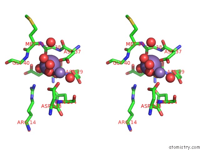
Manganese binding site 2 out of 4 in 5f1m
Go back to
Manganese binding site 2 out
of 4 in the Crystal Structure of Serine/Threonine Phosphatase STP1 From Staphylococcus Aureus
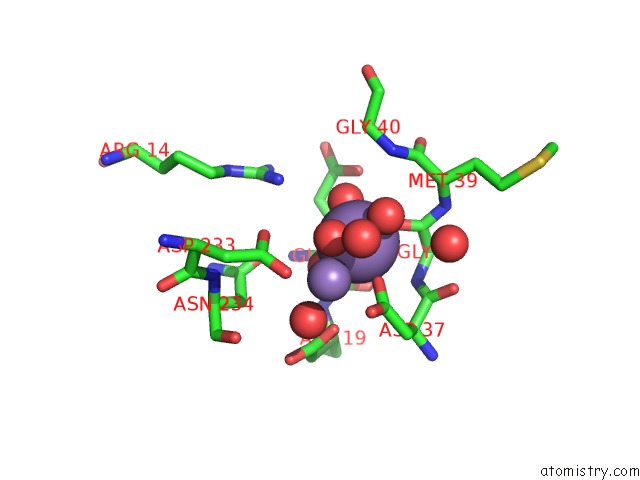
Mono view
Stereo pair view
Mono view
Stereo pair view
A full contact list of Manganese with other atoms in the Mn binding
site number 2 of Crystal Structure of Serine/Threonine Phosphatase STP1 From Staphylococcus Aureus within 5.0Å range:
|
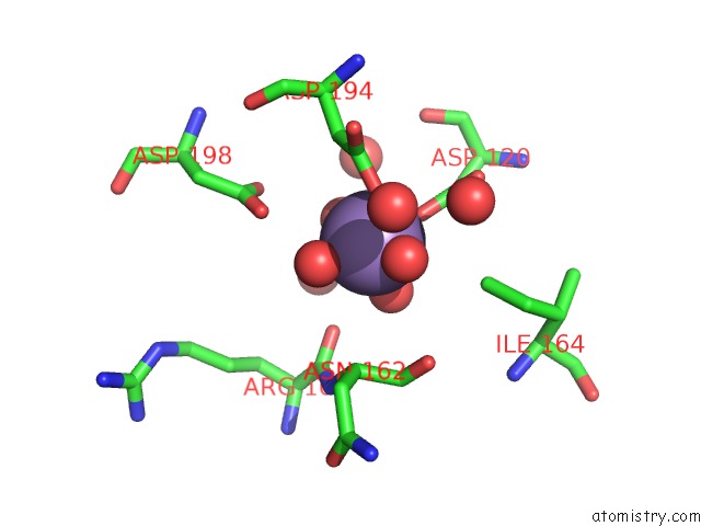
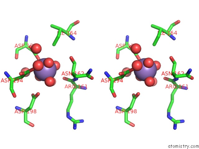
Manganese binding site 3 out of 4 in 5f1m
Go back to
Manganese binding site 3 out
of 4 in the Crystal Structure of Serine/Threonine Phosphatase STP1 From Staphylococcus Aureus
Mono view
Stereo pair view
Mono view
Stereo pair view
A full contact list of Manganese with other atoms in the Mn binding
site number 3 of Crystal Structure of Serine/Threonine Phosphatase STP1 From Staphylococcus Aureus within 5.0Å range:
|
Manganese binding site 4 out of 4 in 5f1m
Go back to
Manganese binding site 4 out
of 4 in the Crystal Structure of Serine/Threonine Phosphatase STP1 From Staphylococcus Aureus
Mono view
Stereo pair view
Mono view
Stereo pair view
A full contact list of Manganese with other atoms in the Mn binding
site number 4 of Crystal Structure of Serine/Threonine Phosphatase STP1 From Staphylococcus Aureus within 5.0Å range:
|
Reference:
W.Zheng,
X.Cai,
M.Xie,
Y.Liang,
T.Wang,
Z.Li.
Structure-Based Identification of A Potent Inhibitor Targeting STP1-Mediated Virulence Regulation in Staphylococcus Aureus Cell Chem Biol V. 23 1002 2016.
ISSN: ESSN 2451-9456
PubMed: 27499528
DOI: 10.1016/J.CHEMBIOL.2016.06.014
Page generated: Sun Oct 6 00:10:36 2024
ISSN: ESSN 2451-9456
PubMed: 27499528
DOI: 10.1016/J.CHEMBIOL.2016.06.014
Last articles
Zn in 9MJ5Zn in 9HNW
Zn in 9G0L
Zn in 9FNE
Zn in 9DZN
Zn in 9E0I
Zn in 9D32
Zn in 9DAK
Zn in 8ZXC
Zn in 8ZUF