Manganese »
PDB 5ekw-5fxv »
5esd »
Manganese in PDB 5esd: Crystal Structure of M. Tuberculosis Mend Bound to Thdp and MN2+
Enzymatic activity of Crystal Structure of M. Tuberculosis Mend Bound to Thdp and MN2+
All present enzymatic activity of Crystal Structure of M. Tuberculosis Mend Bound to Thdp and MN2+:
2.2.1.9;
2.2.1.9;
Protein crystallography data
The structure of Crystal Structure of M. Tuberculosis Mend Bound to Thdp and MN2+, PDB code: 5esd
was solved by
J.M.Johnston,
E.N.M.Jirgis,
G.Bashiri,
E.M.M.Bulloch,
E.N.Baker,
with X-Ray Crystallography technique. A brief refinement statistics is given in the table below:
| Resolution Low / High (Å) | 19.81 / 2.25 |
| Space group | P 21 21 21 |
| Cell size a, b, c (Å), α, β, γ (°) | 98.625, 138.657, 165.386, 90.00, 90.00, 90.00 |
| R / Rfree (%) | 21.1 / 23.5 |
Manganese Binding Sites:
The binding sites of Manganese atom in the Crystal Structure of M. Tuberculosis Mend Bound to Thdp and MN2+
(pdb code 5esd). This binding sites where shown within
5.0 Angstroms radius around Manganese atom.
In total 4 binding sites of Manganese where determined in the Crystal Structure of M. Tuberculosis Mend Bound to Thdp and MN2+, PDB code: 5esd:
Jump to Manganese binding site number: 1; 2; 3; 4;
In total 4 binding sites of Manganese where determined in the Crystal Structure of M. Tuberculosis Mend Bound to Thdp and MN2+, PDB code: 5esd:
Jump to Manganese binding site number: 1; 2; 3; 4;
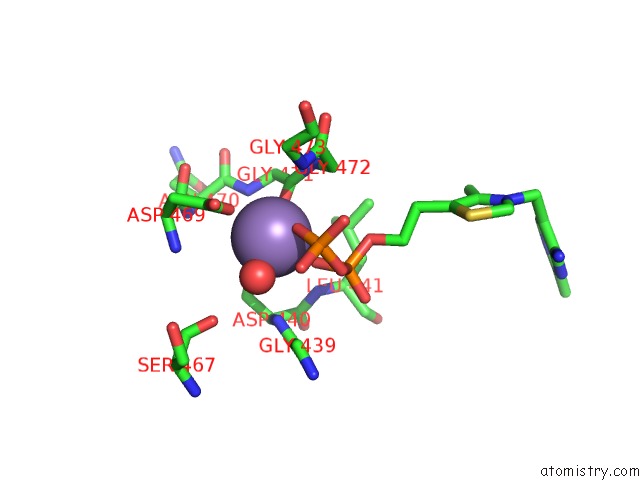
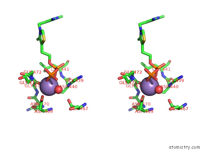
Manganese binding site 1 out of 4 in 5esd
Go back to
Manganese binding site 1 out
of 4 in the Crystal Structure of M. Tuberculosis Mend Bound to Thdp and MN2+
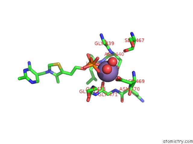
Mono view
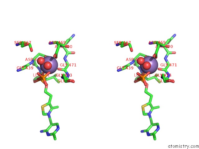
Stereo pair view
Mono view
Stereo pair view
A full contact list of Manganese with other atoms in the Mn binding
site number 1 of Crystal Structure of M. Tuberculosis Mend Bound to Thdp and MN2+ within 5.0Å range:
|
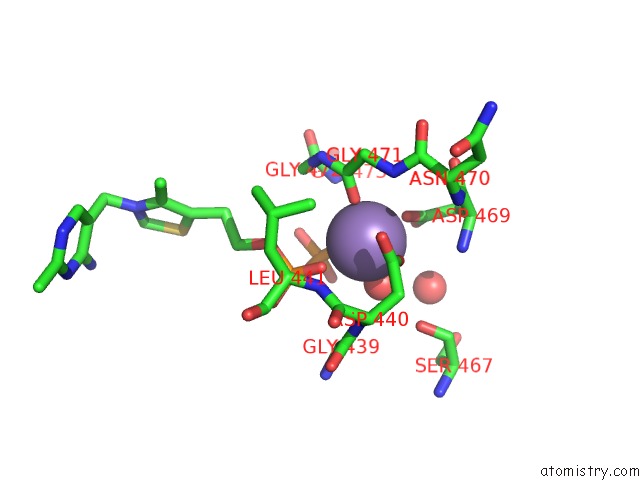
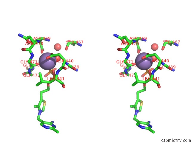
Manganese binding site 2 out of 4 in 5esd
Go back to
Manganese binding site 2 out
of 4 in the Crystal Structure of M. Tuberculosis Mend Bound to Thdp and MN2+
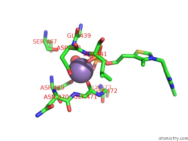
Mono view
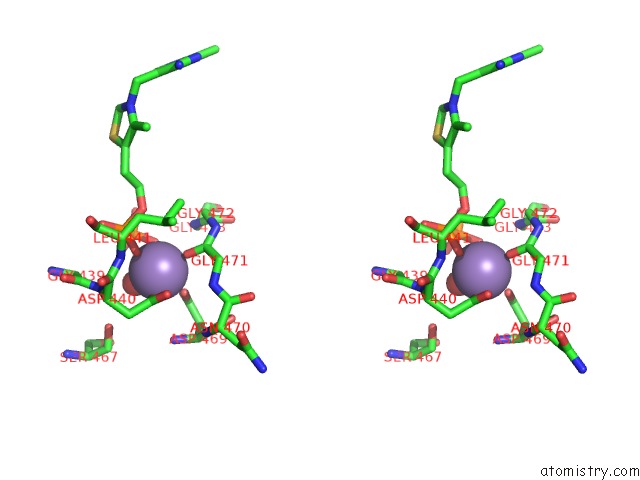
Stereo pair view
Mono view
Stereo pair view
A full contact list of Manganese with other atoms in the Mn binding
site number 2 of Crystal Structure of M. Tuberculosis Mend Bound to Thdp and MN2+ within 5.0Å range:
|
Manganese binding site 3 out of 4 in 5esd
Go back to
Manganese binding site 3 out
of 4 in the Crystal Structure of M. Tuberculosis Mend Bound to Thdp and MN2+
Mono view
Stereo pair view
Mono view
Stereo pair view
A full contact list of Manganese with other atoms in the Mn binding
site number 3 of Crystal Structure of M. Tuberculosis Mend Bound to Thdp and MN2+ within 5.0Å range:
|
Manganese binding site 4 out of 4 in 5esd
Go back to
Manganese binding site 4 out
of 4 in the Crystal Structure of M. Tuberculosis Mend Bound to Thdp and MN2+
Mono view
Stereo pair view
Mono view
Stereo pair view
A full contact list of Manganese with other atoms in the Mn binding
site number 4 of Crystal Structure of M. Tuberculosis Mend Bound to Thdp and MN2+ within 5.0Å range:
|
Reference:
E.N.Jirgis,
G.Bashiri,
E.M.Bulloch,
J.M.Johnston,
E.N.Baker.
Structural Views Along the Mycobacterium Tuberculosis Mend Reaction Pathway Illuminate Key Aspects of Thiamin Diphosphate-Dependent Enzyme Mechanisms. Structure V. 24 1167 2016.
ISSN: ISSN 0969-2126
PubMed: 27291649
DOI: 10.1016/J.STR.2016.04.018
Page generated: Sun Oct 6 00:09:19 2024
ISSN: ISSN 0969-2126
PubMed: 27291649
DOI: 10.1016/J.STR.2016.04.018
Last articles
Zn in 9JYWZn in 9IR4
Zn in 9IR3
Zn in 9GMX
Zn in 9GMW
Zn in 9JEJ
Zn in 9ERF
Zn in 9ERE
Zn in 9EGV
Zn in 9EGW