Manganese »
PDB 5cdn-5dcb »
5cna »
Manganese in PDB 5cna: Refined Structure of Concanavalin A Complexed with Alpha- Methyl-D-Mannopyranoside at 2.0 Angstroms Resolution and Comparison with the Saccharide-Free Structure
Protein crystallography data
The structure of Refined Structure of Concanavalin A Complexed with Alpha- Methyl-D-Mannopyranoside at 2.0 Angstroms Resolution and Comparison with the Saccharide-Free Structure, PDB code: 5cna
was solved by
J.H.Naismith,
C.Emmerich,
J.Habash,
S.J.Harrop,
J.R.Helliwell,
W.N.Hunter,
J.Raftery,
A.J.Kalb(Gilboa),
J.Yariv,
with X-Ray Crystallography technique. A brief refinement statistics is given in the table below:
| Resolution Low / High (Å) | 8.00 / 2.00 |
| Space group | P 21 21 21 |
| Cell size a, b, c (Å), α, β, γ (°) | 123.700, 128.600, 67.200, 90.00, 90.00, 90.00 |
| R / Rfree (%) | 19.9 / n/a |
Other elements in 5cna:
The structure of Refined Structure of Concanavalin A Complexed with Alpha- Methyl-D-Mannopyranoside at 2.0 Angstroms Resolution and Comparison with the Saccharide-Free Structure also contains other interesting chemical elements:
| Chlorine | (Cl) | 1 atom |
| Calcium | (Ca) | 4 atoms |
Manganese Binding Sites:
The binding sites of Manganese atom in the Refined Structure of Concanavalin A Complexed with Alpha- Methyl-D-Mannopyranoside at 2.0 Angstroms Resolution and Comparison with the Saccharide-Free Structure
(pdb code 5cna). This binding sites where shown within
5.0 Angstroms radius around Manganese atom.
In total 4 binding sites of Manganese where determined in the Refined Structure of Concanavalin A Complexed with Alpha- Methyl-D-Mannopyranoside at 2.0 Angstroms Resolution and Comparison with the Saccharide-Free Structure, PDB code: 5cna:
Jump to Manganese binding site number: 1; 2; 3; 4;
In total 4 binding sites of Manganese where determined in the Refined Structure of Concanavalin A Complexed with Alpha- Methyl-D-Mannopyranoside at 2.0 Angstroms Resolution and Comparison with the Saccharide-Free Structure, PDB code: 5cna:
Jump to Manganese binding site number: 1; 2; 3; 4;
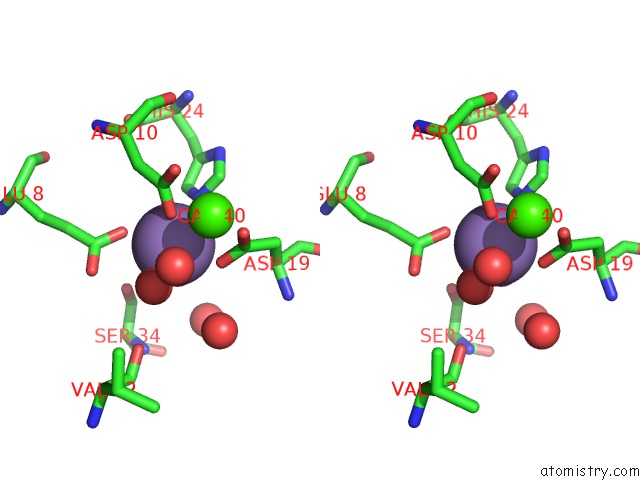
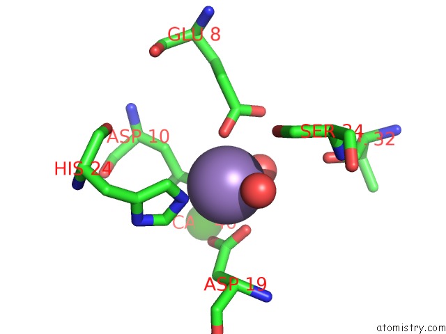
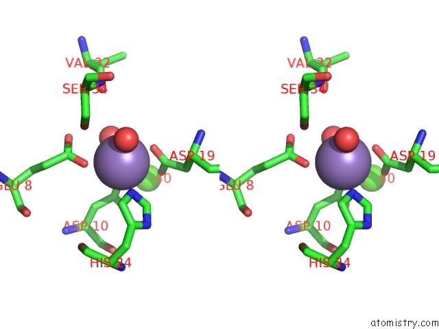
Manganese binding site 1 out of 4 in 5cna
Go back to
Manganese binding site 1 out
of 4 in the Refined Structure of Concanavalin A Complexed with Alpha- Methyl-D-Mannopyranoside at 2.0 Angstroms Resolution and Comparison with the Saccharide-Free Structure
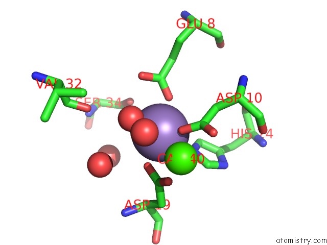
Mono view
Stereo pair view
Mono view
Stereo pair view
A full contact list of Manganese with other atoms in the Mn binding
site number 1 of Refined Structure of Concanavalin A Complexed with Alpha- Methyl-D-Mannopyranoside at 2.0 Angstroms Resolution and Comparison with the Saccharide-Free Structure within 5.0Å range:
|
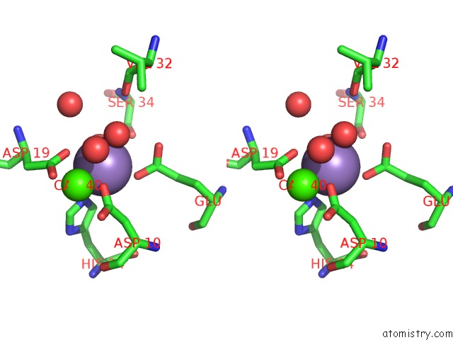
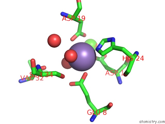
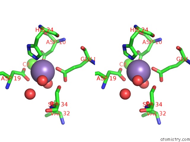
Manganese binding site 2 out of 4 in 5cna
Go back to
Manganese binding site 2 out
of 4 in the Refined Structure of Concanavalin A Complexed with Alpha- Methyl-D-Mannopyranoside at 2.0 Angstroms Resolution and Comparison with the Saccharide-Free Structure
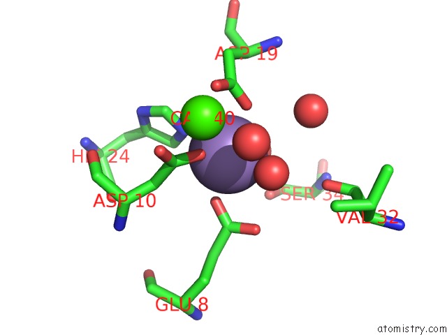
Mono view
Stereo pair view
Mono view
Stereo pair view
A full contact list of Manganese with other atoms in the Mn binding
site number 2 of Refined Structure of Concanavalin A Complexed with Alpha- Methyl-D-Mannopyranoside at 2.0 Angstroms Resolution and Comparison with the Saccharide-Free Structure within 5.0Å range:
|
Manganese binding site 3 out of 4 in 5cna
Go back to
Manganese binding site 3 out
of 4 in the Refined Structure of Concanavalin A Complexed with Alpha- Methyl-D-Mannopyranoside at 2.0 Angstroms Resolution and Comparison with the Saccharide-Free Structure
Mono view
Stereo pair view
Mono view
Stereo pair view
A full contact list of Manganese with other atoms in the Mn binding
site number 3 of Refined Structure of Concanavalin A Complexed with Alpha- Methyl-D-Mannopyranoside at 2.0 Angstroms Resolution and Comparison with the Saccharide-Free Structure within 5.0Å range:
|
Manganese binding site 4 out of 4 in 5cna
Go back to
Manganese binding site 4 out
of 4 in the Refined Structure of Concanavalin A Complexed with Alpha- Methyl-D-Mannopyranoside at 2.0 Angstroms Resolution and Comparison with the Saccharide-Free Structure
Mono view
Stereo pair view
Mono view
Stereo pair view
A full contact list of Manganese with other atoms in the Mn binding
site number 4 of Refined Structure of Concanavalin A Complexed with Alpha- Methyl-D-Mannopyranoside at 2.0 Angstroms Resolution and Comparison with the Saccharide-Free Structure within 5.0Å range:
|
Reference:
J.H.Naismith,
C.Emmerich,
J.Habash,
S.J.Harrop,
J.R.Helliwell,
W.N.Hunter,
J.Raftery,
A.J.Kalb,
J.Yariv.
Refined Structure of Concanavalin A Complexed with Methyl Alpha-D-Mannopyranoside at 2.0 A Resolution and Comparison with the Saccharide-Free Structure. Acta Crystallogr.,Sect.D V. 50 847 1994.
ISSN: ISSN 0907-4449
PubMed: 15299352
DOI: 10.1107/S0907444994005287
Page generated: Sat Oct 5 23:47:45 2024
ISSN: ISSN 0907-4449
PubMed: 15299352
DOI: 10.1107/S0907444994005287
Last articles
Zn in 9J0NZn in 9J0O
Zn in 9J0P
Zn in 9FJX
Zn in 9EKB
Zn in 9C0F
Zn in 9CAH
Zn in 9CH0
Zn in 9CH3
Zn in 9CH1