Manganese »
PDB 5cdn-5dcb »
5cet »
Manganese in PDB 5cet: Crystal Structure of RV2837C
Enzymatic activity of Crystal Structure of RV2837C
All present enzymatic activity of Crystal Structure of RV2837C:
3.1.3.7;
3.1.3.7;
Protein crystallography data
The structure of Crystal Structure of RV2837C, PDB code: 5cet
was solved by
F.Wang,
Q.He,
D.Zhu,
S.Liu,
L.Gu,
with X-Ray Crystallography technique. A brief refinement statistics is given in the table below:
| Resolution Low / High (Å) | 37.26 / 2.00 |
| Space group | P 21 21 2 |
| Cell size a, b, c (Å), α, β, γ (°) | 58.864, 98.614, 55.143, 90.00, 90.00, 90.00 |
| R / Rfree (%) | 20.1 / 23.6 |
Manganese Binding Sites:
The binding sites of Manganese atom in the Crystal Structure of RV2837C
(pdb code 5cet). This binding sites where shown within
5.0 Angstroms radius around Manganese atom.
In total 2 binding sites of Manganese where determined in the Crystal Structure of RV2837C, PDB code: 5cet:
Jump to Manganese binding site number: 1; 2;
In total 2 binding sites of Manganese where determined in the Crystal Structure of RV2837C, PDB code: 5cet:
Jump to Manganese binding site number: 1; 2;
Manganese binding site 1 out of 2 in 5cet
Go back to
Manganese binding site 1 out
of 2 in the Crystal Structure of RV2837C
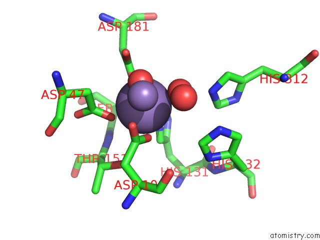
Mono view
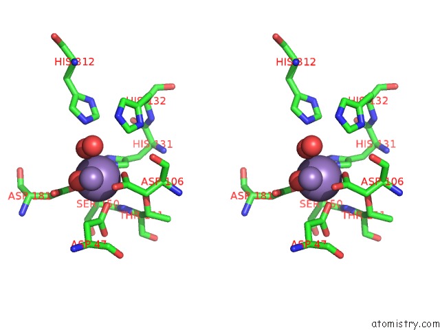
Stereo pair view
Mono view
Stereo pair view
A full contact list of Manganese with other atoms in the Mn binding
site number 1 of Crystal Structure of RV2837C within 5.0Å range:
|
Manganese binding site 2 out of 2 in 5cet
Go back to
Manganese binding site 2 out
of 2 in the Crystal Structure of RV2837C
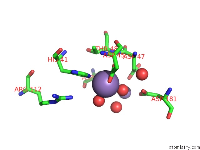
Mono view
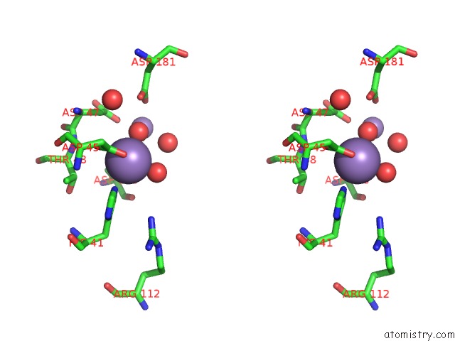
Stereo pair view
Mono view
Stereo pair view
A full contact list of Manganese with other atoms in the Mn binding
site number 2 of Crystal Structure of RV2837C within 5.0Å range:
|
Reference:
Q.He,
F.Wang,
S.Liu,
D.Zhu,
H.Cong,
F.Gao,
B.Li,
H.Wang,
Z.Lin,
J.Liao,
L.Gu.
Structural and Biochemical Insight Into the Mechanism of RV2837C From Mycobacterium Tuberculosis As A C-Di-Nmp Phosphodiesterase J.Biol.Chem. V. 291 3668 2016.
ISSN: ESSN 1083-351X
PubMed: 26668313
DOI: 10.1074/JBC.M115.699801
Page generated: Sat Oct 5 23:45:18 2024
ISSN: ESSN 1083-351X
PubMed: 26668313
DOI: 10.1074/JBC.M115.699801
Last articles
Cl in 7TNKCl in 7TNJ
Cl in 7TNH
Cl in 7TNF
Cl in 7TND
Cl in 7TN8
Cl in 7TNC
Cl in 7TN7
Cl in 7TN4
Cl in 7TN6