Manganese »
PDB 5b4c-5cdm »
5c4e »
Manganese in PDB 5c4e: Crystal Structure of Gtb + Udp-Glc + H-Antigen Acceptor
Enzymatic activity of Crystal Structure of Gtb + Udp-Glc + H-Antigen Acceptor
All present enzymatic activity of Crystal Structure of Gtb + Udp-Glc + H-Antigen Acceptor:
2.4.1.37; 2.4.1.40;
2.4.1.37; 2.4.1.40;
Protein crystallography data
The structure of Crystal Structure of Gtb + Udp-Glc + H-Antigen Acceptor, PDB code: 5c4e
was solved by
S.Gagnon,
P.Meloncelli,
R.B.Zheng,
O.Haji-Ghassemi,
A.R.Johal,
S.Borisova,
T.L.Lowary,
S.V.Evans,
with X-Ray Crystallography technique. A brief refinement statistics is given in the table below:
| Resolution Low / High (Å) | 74.83 / 1.55 |
| Space group | C 2 2 21 |
| Cell size a, b, c (Å), α, β, γ (°) | 52.640, 149.670, 79.460, 90.00, 90.00, 90.00 |
| R / Rfree (%) | 17.3 / 19.8 |
Manganese Binding Sites:
The binding sites of Manganese atom in the Crystal Structure of Gtb + Udp-Glc + H-Antigen Acceptor
(pdb code 5c4e). This binding sites where shown within
5.0 Angstroms radius around Manganese atom.
In total only one binding site of Manganese was determined in the Crystal Structure of Gtb + Udp-Glc + H-Antigen Acceptor, PDB code: 5c4e:
In total only one binding site of Manganese was determined in the Crystal Structure of Gtb + Udp-Glc + H-Antigen Acceptor, PDB code: 5c4e:
Manganese binding site 1 out of 1 in 5c4e
Go back to
Manganese binding site 1 out
of 1 in the Crystal Structure of Gtb + Udp-Glc + H-Antigen Acceptor
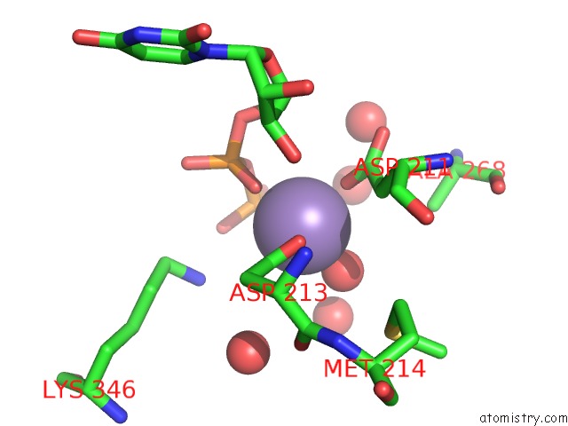
Mono view
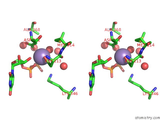
Stereo pair view
Mono view
Stereo pair view
A full contact list of Manganese with other atoms in the Mn binding
site number 1 of Crystal Structure of Gtb + Udp-Glc + H-Antigen Acceptor within 5.0Å range:
|
Reference:
S.M.Gagnon,
P.J.Meloncelli,
R.B.Zheng,
O.Haji-Ghassemi,
A.R.Johal,
S.N.Borisova,
T.L.Lowary,
S.V.Evans.
High Resolution Structures of the Human Abo(H) Blood Group Enzymes in Complex with Donor Analogs Reveal That the Enzymes Utilize Multiple Donor Conformations to Bind Substrates in A Stepwise Manner. J.Biol.Chem. V. 290 27040 2015.
ISSN: ESSN 1083-351X
PubMed: 26374898
DOI: 10.1074/JBC.M115.682401
Page generated: Sat Oct 5 23:43:37 2024
ISSN: ESSN 1083-351X
PubMed: 26374898
DOI: 10.1074/JBC.M115.682401
Last articles
Zn in 9J0NZn in 9J0O
Zn in 9J0P
Zn in 9FJX
Zn in 9EKB
Zn in 9C0F
Zn in 9CAH
Zn in 9CH0
Zn in 9CH3
Zn in 9CH1