Manganese »
PDB 5b4c-5cdm »
5b4c »
Manganese in PDB 5b4c: Crystal Structure of H10N Mutant of Lpxh with Manganese
Enzymatic activity of Crystal Structure of H10N Mutant of Lpxh with Manganese
All present enzymatic activity of Crystal Structure of H10N Mutant of Lpxh with Manganese:
3.6.1.54;
3.6.1.54;
Protein crystallography data
The structure of Crystal Structure of H10N Mutant of Lpxh with Manganese, PDB code: 5b4c
was solved by
C.Okada,
H.Wakabayashi,
M.Yao,
I.Tanaka,
with X-Ray Crystallography technique. A brief refinement statistics is given in the table below:
| Resolution Low / High (Å) | 49.15 / 1.96 |
| Space group | P 21 21 21 |
| Cell size a, b, c (Å), α, β, γ (°) | 53.622, 97.999, 113.631, 90.00, 90.00, 90.00 |
| R / Rfree (%) | 17.1 / 21.4 |
Manganese Binding Sites:
The binding sites of Manganese atom in the Crystal Structure of H10N Mutant of Lpxh with Manganese
(pdb code 5b4c). This binding sites where shown within
5.0 Angstroms radius around Manganese atom.
In total 2 binding sites of Manganese where determined in the Crystal Structure of H10N Mutant of Lpxh with Manganese, PDB code: 5b4c:
Jump to Manganese binding site number: 1; 2;
In total 2 binding sites of Manganese where determined in the Crystal Structure of H10N Mutant of Lpxh with Manganese, PDB code: 5b4c:
Jump to Manganese binding site number: 1; 2;
Manganese binding site 1 out of 2 in 5b4c
Go back to
Manganese binding site 1 out
of 2 in the Crystal Structure of H10N Mutant of Lpxh with Manganese
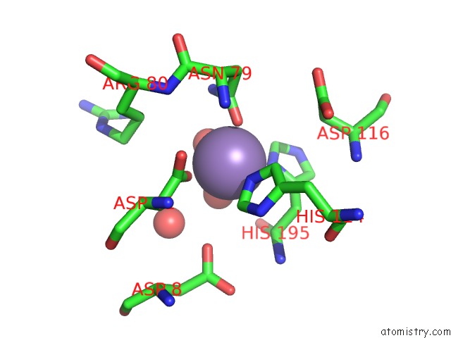
Mono view
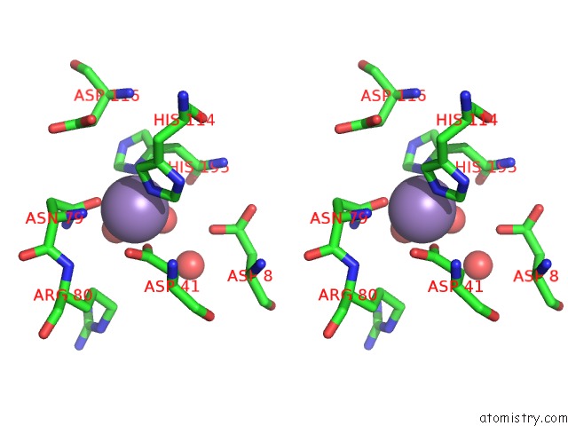
Stereo pair view
Mono view
Stereo pair view
A full contact list of Manganese with other atoms in the Mn binding
site number 1 of Crystal Structure of H10N Mutant of Lpxh with Manganese within 5.0Å range:
|
Manganese binding site 2 out of 2 in 5b4c
Go back to
Manganese binding site 2 out
of 2 in the Crystal Structure of H10N Mutant of Lpxh with Manganese
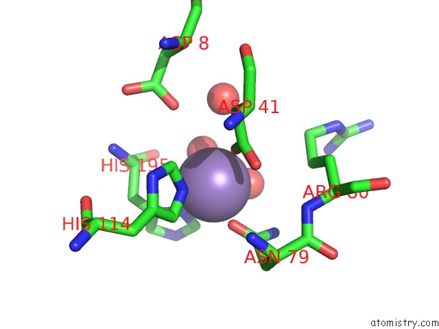
Mono view
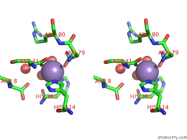
Stereo pair view
Mono view
Stereo pair view
A full contact list of Manganese with other atoms in the Mn binding
site number 2 of Crystal Structure of H10N Mutant of Lpxh with Manganese within 5.0Å range:
|
Reference:
C.Okada,
H.Wakabayashi,
M.Kobayashi,
A.Shinoda,
I.Tanaka,
M.Yao.
Crystal Structures of the Udp-Diacylglucosamine Pyrophosphohydrase Lpxh From Pseudomonas Aeruginosa Sci Rep V. 6 32822 2016.
ISSN: ESSN 2045-2322
PubMed: 27609419
DOI: 10.1038/SREP32822
Page generated: Sat Oct 5 23:37:53 2024
ISSN: ESSN 2045-2322
PubMed: 27609419
DOI: 10.1038/SREP32822
Last articles
Zn in 9J0NZn in 9J0O
Zn in 9J0P
Zn in 9FJX
Zn in 9EKB
Zn in 9C0F
Zn in 9CAH
Zn in 9CH0
Zn in 9CH3
Zn in 9CH1