Manganese »
PDB 4z8b-5a56 »
5a55 »
Manganese in PDB 5a55: The Native Structure of GH101 From Streptococcus Pneumoniae TIGR4
Enzymatic activity of The Native Structure of GH101 From Streptococcus Pneumoniae TIGR4
All present enzymatic activity of The Native Structure of GH101 From Streptococcus Pneumoniae TIGR4:
3.2.1.97;
3.2.1.97;
Protein crystallography data
The structure of The Native Structure of GH101 From Streptococcus Pneumoniae TIGR4, PDB code: 5a55
was solved by
K.J.Gregg,
M.D.L.Suits,
L.Deng,
D.J.Vocadlo,
A.B.Boraston,
with X-Ray Crystallography technique. A brief refinement statistics is given in the table below:
| Resolution Low / High (Å) | 89.09 / 1.85 |
| Space group | P 1 21 1 |
| Cell size a, b, c (Å), α, β, γ (°) | 76.260, 89.130, 88.570, 90.00, 110.91, 90.00 |
| R / Rfree (%) | 14.3 / 17.9 |
Other elements in 5a55:
The structure of The Native Structure of GH101 From Streptococcus Pneumoniae TIGR4 also contains other interesting chemical elements:
| Calcium | (Ca) | 4 atoms |
Manganese Binding Sites:
The binding sites of Manganese atom in the The Native Structure of GH101 From Streptococcus Pneumoniae TIGR4
(pdb code 5a55). This binding sites where shown within
5.0 Angstroms radius around Manganese atom.
In total only one binding site of Manganese was determined in the The Native Structure of GH101 From Streptococcus Pneumoniae TIGR4, PDB code: 5a55:
In total only one binding site of Manganese was determined in the The Native Structure of GH101 From Streptococcus Pneumoniae TIGR4, PDB code: 5a55:
Manganese binding site 1 out of 1 in 5a55
Go back to
Manganese binding site 1 out
of 1 in the The Native Structure of GH101 From Streptococcus Pneumoniae TIGR4
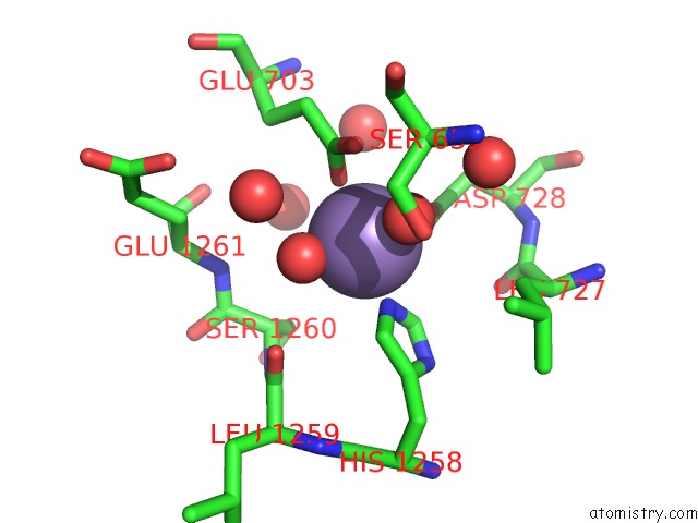
Mono view
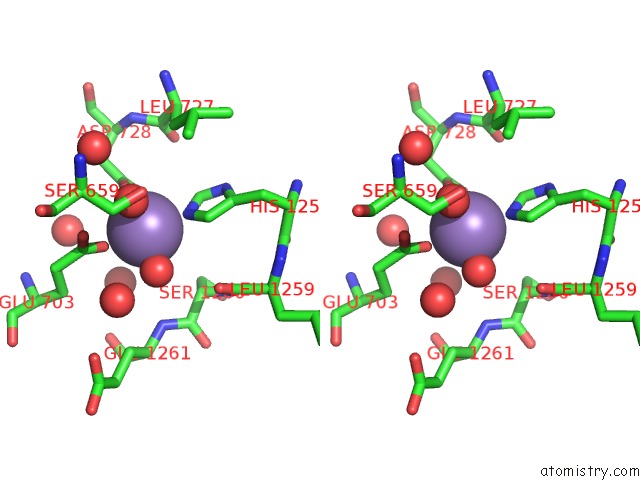
Stereo pair view
Mono view
Stereo pair view
A full contact list of Manganese with other atoms in the Mn binding
site number 1 of The Native Structure of GH101 From Streptococcus Pneumoniae TIGR4 within 5.0Å range:
|
Reference:
K.J.Gregg,
M.D.L.Suits,
L.Deng,
D.J.Vocadlo,
A.B.Boraston.
Structural Analysis of A Family 101 Glycoside Hydrolase in Complex with Carbohydrates Reveals Insights Into Its Mechanism. J.Biol.Chem. V. 290 25657 2015.
ISSN: ISSN 0021-9258
PubMed: 26304114
DOI: 10.1074/JBC.M115.680470
Page generated: Sat Oct 5 23:20:52 2024
ISSN: ISSN 0021-9258
PubMed: 26304114
DOI: 10.1074/JBC.M115.680470
Last articles
Zn in 9J0NZn in 9J0O
Zn in 9J0P
Zn in 9FJX
Zn in 9EKB
Zn in 9C0F
Zn in 9CAH
Zn in 9CH0
Zn in 9CH3
Zn in 9CH1