Manganese »
PDB 4z8b-5a56 »
5a29 »
Manganese in PDB 5a29: Family 2 Pectate Lyase From Vibrio Vulnificus
Protein crystallography data
The structure of Family 2 Pectate Lyase From Vibrio Vulnificus, PDB code: 5a29
was solved by
R.Mclean,
J.K.Hobbs,
M.D.Suits,
S.Tuomivaara,
D.Jones,
A.B.Boraston,
D.W.Abbott,
with X-Ray Crystallography technique. A brief refinement statistics is given in the table below:
| Resolution Low / High (Å) | 122.20 / 1.90 |
| Space group | P 65 |
| Cell size a, b, c (Å), α, β, γ (°) | 141.102, 141.102, 72.426, 90.00, 90.00, 120.00 |
| R / Rfree (%) | 15.6 / 19.7 |
Manganese Binding Sites:
The binding sites of Manganese atom in the Family 2 Pectate Lyase From Vibrio Vulnificus
(pdb code 5a29). This binding sites where shown within
5.0 Angstroms radius around Manganese atom.
In total only one binding site of Manganese was determined in the Family 2 Pectate Lyase From Vibrio Vulnificus, PDB code: 5a29:
In total only one binding site of Manganese was determined in the Family 2 Pectate Lyase From Vibrio Vulnificus, PDB code: 5a29:
Manganese binding site 1 out of 1 in 5a29
Go back to
Manganese binding site 1 out
of 1 in the Family 2 Pectate Lyase From Vibrio Vulnificus
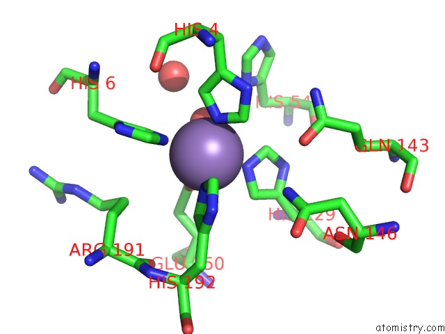
Mono view
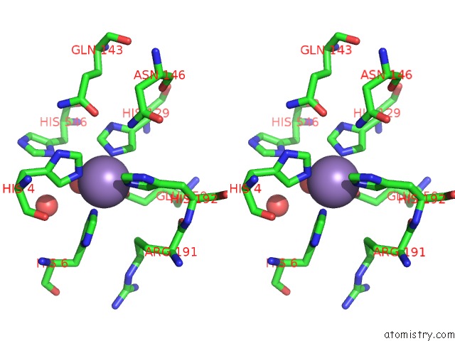
Stereo pair view
Mono view
Stereo pair view
A full contact list of Manganese with other atoms in the Mn binding
site number 1 of Family 2 Pectate Lyase From Vibrio Vulnificus within 5.0Å range:
|
Reference:
R.Mclean,
J.K.Hobbs,
M.D.Suits,
S.T.Tuomivaara,
D.Jones,
A.B.Boraston,
D.W.Abbott.
Functional Analyses of Resurrected and Contemporary Enzymes Illuminate An Evolutionary Path For the Emergence of Exolysis in Polysaccharide Lyase Family 2. J.Biol.Chem. V. 290 21231 2015.
ISSN: ISSN 0021-9258
PubMed: 26160170
DOI: 10.1074/JBC.M115.664847
Page generated: Sat Oct 5 23:19:43 2024
ISSN: ISSN 0021-9258
PubMed: 26160170
DOI: 10.1074/JBC.M115.664847
Last articles
Cl in 5U4QCl in 5U4F
Cl in 5U4E
Cl in 5U4M
Cl in 5U4G
Cl in 5U2G
Cl in 5U4D
Cl in 5U4A
Cl in 5U3Q
Cl in 5U49