Manganese »
PDB 4z8b-5a56 »
5a07 »
Manganese in PDB 5a07: X-Ray Structure of the Mannosyltransferase KTR4P From S. Cerevisiae in Complex with Gdp
Protein crystallography data
The structure of X-Ray Structure of the Mannosyltransferase KTR4P From S. Cerevisiae in Complex with Gdp, PDB code: 5a07
was solved by
D.D.D.Possner,
J.E.Guy,
with X-Ray Crystallography technique. A brief refinement statistics is given in the table below:
| Resolution Low / High (Å) | 86.79 / 1.90 |
| Space group | P 21 21 21 |
| Cell size a, b, c (Å), α, β, γ (°) | 61.215, 102.621, 162.651, 90.00, 90.00, 90.00 |
| R / Rfree (%) | 15.627 / 19.132 |
Manganese Binding Sites:
The binding sites of Manganese atom in the X-Ray Structure of the Mannosyltransferase KTR4P From S. Cerevisiae in Complex with Gdp
(pdb code 5a07). This binding sites where shown within
5.0 Angstroms radius around Manganese atom.
In total 2 binding sites of Manganese where determined in the X-Ray Structure of the Mannosyltransferase KTR4P From S. Cerevisiae in Complex with Gdp, PDB code: 5a07:
Jump to Manganese binding site number: 1; 2;
In total 2 binding sites of Manganese where determined in the X-Ray Structure of the Mannosyltransferase KTR4P From S. Cerevisiae in Complex with Gdp, PDB code: 5a07:
Jump to Manganese binding site number: 1; 2;
Manganese binding site 1 out of 2 in 5a07
Go back to
Manganese binding site 1 out
of 2 in the X-Ray Structure of the Mannosyltransferase KTR4P From S. Cerevisiae in Complex with Gdp
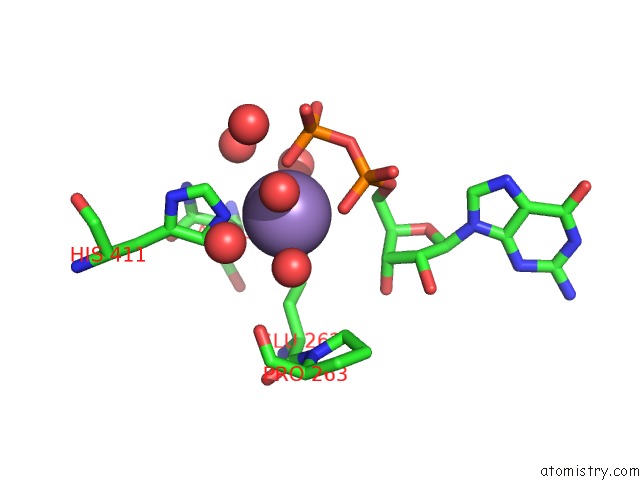
Mono view
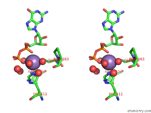
Stereo pair view
Mono view
Stereo pair view
A full contact list of Manganese with other atoms in the Mn binding
site number 1 of X-Ray Structure of the Mannosyltransferase KTR4P From S. Cerevisiae in Complex with Gdp within 5.0Å range:
|
Manganese binding site 2 out of 2 in 5a07
Go back to
Manganese binding site 2 out
of 2 in the X-Ray Structure of the Mannosyltransferase KTR4P From S. Cerevisiae in Complex with Gdp
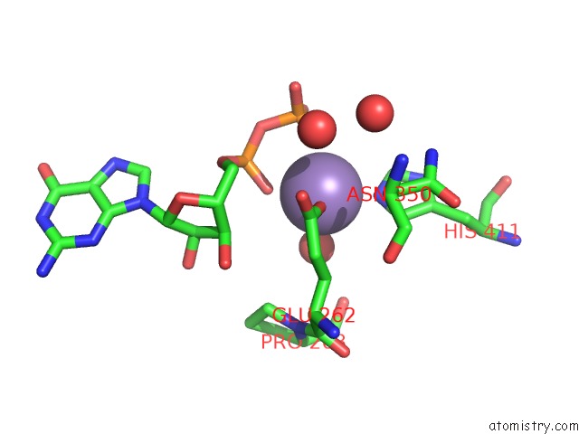
Mono view
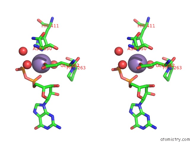
Stereo pair view
Mono view
Stereo pair view
A full contact list of Manganese with other atoms in the Mn binding
site number 2 of X-Ray Structure of the Mannosyltransferase KTR4P From S. Cerevisiae in Complex with Gdp within 5.0Å range:
|
Reference:
D.D.D.Possner,
M.Claesson,
J.E.Guy.
Structure of the Glycosyltransferase KTR4P From Saccharomyces Cerevisiae Plos One V. 10 36239 2015.
ISSN: ISSN 1932-6203
PubMed: 26296208
DOI: 10.1371/JOURNAL.PONE.013623
Page generated: Sat Oct 5 23:17:38 2024
ISSN: ISSN 1932-6203
PubMed: 26296208
DOI: 10.1371/JOURNAL.PONE.013623
Last articles
Zn in 9J0NZn in 9J0O
Zn in 9J0P
Zn in 9FJX
Zn in 9EKB
Zn in 9C0F
Zn in 9CAH
Zn in 9CH0
Zn in 9CH3
Zn in 9CH1