Manganese »
PDB 4z8b-5a56 »
4z8b »
Manganese in PDB 4z8b: Crystal Structure of A Dgl Mutant - H51G H131N
Protein crystallography data
The structure of Crystal Structure of A Dgl Mutant - H51G H131N, PDB code: 4z8b
was solved by
S.Zamora-Caballero,
A.Perez,
L.Sanz,
J.Bravo,
J.J.Calvete,
with X-Ray Crystallography technique. A brief refinement statistics is given in the table below:
| Resolution Low / High (Å) | 45.67 / 1.95 |
| Space group | I 2 2 2 |
| Cell size a, b, c (Å), α, β, γ (°) | 62.350, 67.080, 108.620, 90.00, 90.00, 90.00 |
| R / Rfree (%) | 20.1 / 24.3 |
Other elements in 4z8b:
The structure of Crystal Structure of A Dgl Mutant - H51G H131N also contains other interesting chemical elements:
| Bromine | (Br) | 1 atom |
| Calcium | (Ca) | 1 atom |
| Chlorine | (Cl) | 1 atom |
Manganese Binding Sites:
The binding sites of Manganese atom in the Crystal Structure of A Dgl Mutant - H51G H131N
(pdb code 4z8b). This binding sites where shown within
5.0 Angstroms radius around Manganese atom.
In total only one binding site of Manganese was determined in the Crystal Structure of A Dgl Mutant - H51G H131N, PDB code: 4z8b:
In total only one binding site of Manganese was determined in the Crystal Structure of A Dgl Mutant - H51G H131N, PDB code: 4z8b:
Manganese binding site 1 out of 1 in 4z8b
Go back to
Manganese binding site 1 out
of 1 in the Crystal Structure of A Dgl Mutant - H51G H131N
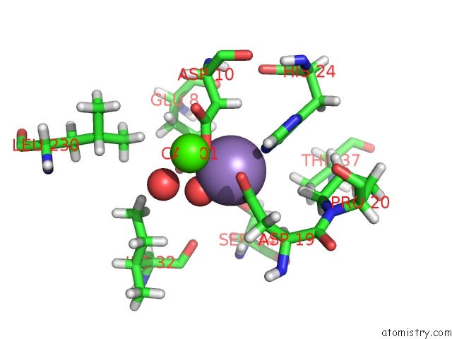
Mono view
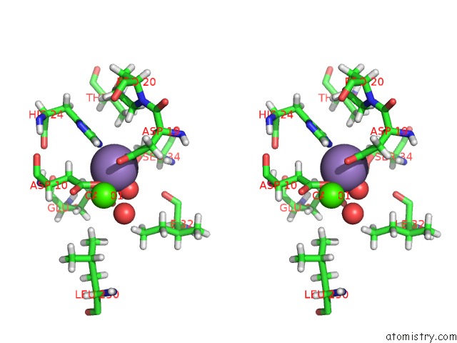
Stereo pair view
Mono view
Stereo pair view
A full contact list of Manganese with other atoms in the Mn binding
site number 1 of Crystal Structure of A Dgl Mutant - H51G H131N within 5.0Å range:
|
Reference:
S.Zamora-Caballero,
A.Perez,
L.Sanz,
J.Bravo,
J.J.Calvete.
Quaternary Structure of Dioclea Grandiflora Lectin Assessed By Equilibrium Sedimentation and Crystallographic Analysis of Recombinant Mutants. Febs Lett. V. 589 2290 2015.
ISSN: ISSN 0014-5793
PubMed: 26226421
DOI: 10.1016/J.FEBSLET.2015.07.020
Page generated: Sat Oct 5 23:12:12 2024
ISSN: ISSN 0014-5793
PubMed: 26226421
DOI: 10.1016/J.FEBSLET.2015.07.020
Last articles
Zn in 9MJ5Zn in 9HNW
Zn in 9G0L
Zn in 9FNE
Zn in 9DZN
Zn in 9E0I
Zn in 9D32
Zn in 9DAK
Zn in 8ZXC
Zn in 8ZUF