Manganese »
PDB 4wl8-4xwt »
4xsl »
Manganese in PDB 4xsl: Crystal Strcutre of D-Tagatose 3-Epimerase C66S From Pseudomonas Cichorii in Complex with Glycerol
Protein crystallography data
The structure of Crystal Strcutre of D-Tagatose 3-Epimerase C66S From Pseudomonas Cichorii in Complex with Glycerol, PDB code: 4xsl
was solved by
H.Yoshida,
A.Yoshihara,
T.Ishii,
K.Izumori,
S.Kamitori,
with X-Ray Crystallography technique. A brief refinement statistics is given in the table below:
| Resolution Low / High (Å) | 32.07 / 1.60 |
| Space group | P 1 21 1 |
| Cell size a, b, c (Å), α, β, γ (°) | 52.271, 124.552, 93.319, 90.00, 98.74, 90.00 |
| R / Rfree (%) | 17.8 / 20.5 |
Manganese Binding Sites:
The binding sites of Manganese atom in the Crystal Strcutre of D-Tagatose 3-Epimerase C66S From Pseudomonas Cichorii in Complex with Glycerol
(pdb code 4xsl). This binding sites where shown within
5.0 Angstroms radius around Manganese atom.
In total 4 binding sites of Manganese where determined in the Crystal Strcutre of D-Tagatose 3-Epimerase C66S From Pseudomonas Cichorii in Complex with Glycerol, PDB code: 4xsl:
Jump to Manganese binding site number: 1; 2; 3; 4;
In total 4 binding sites of Manganese where determined in the Crystal Strcutre of D-Tagatose 3-Epimerase C66S From Pseudomonas Cichorii in Complex with Glycerol, PDB code: 4xsl:
Jump to Manganese binding site number: 1; 2; 3; 4;
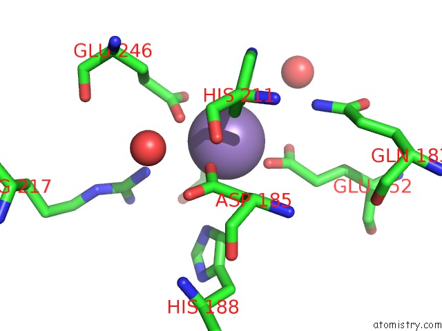
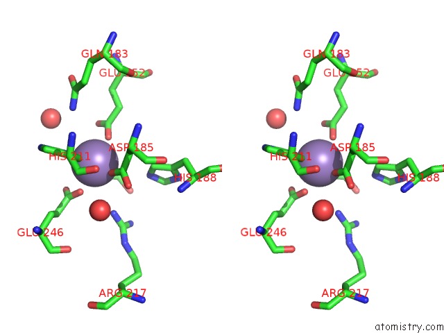
Manganese binding site 1 out of 4 in 4xsl
Go back to
Manganese binding site 1 out
of 4 in the Crystal Strcutre of D-Tagatose 3-Epimerase C66S From Pseudomonas Cichorii in Complex with Glycerol
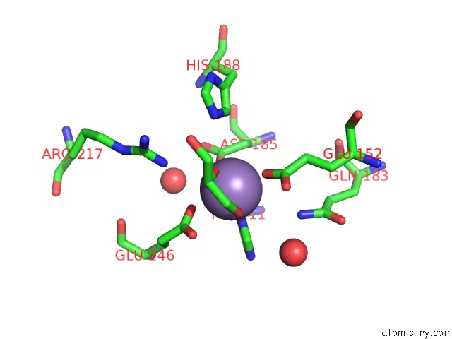
Mono view
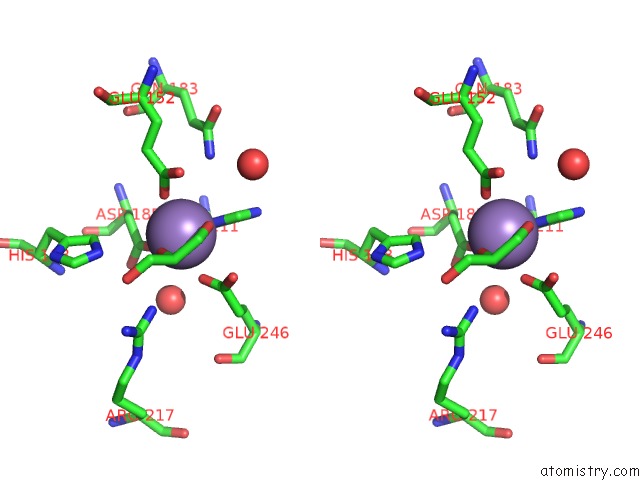
Stereo pair view
Mono view
Stereo pair view
A full contact list of Manganese with other atoms in the Mn binding
site number 1 of Crystal Strcutre of D-Tagatose 3-Epimerase C66S From Pseudomonas Cichorii in Complex with Glycerol within 5.0Å range:
|
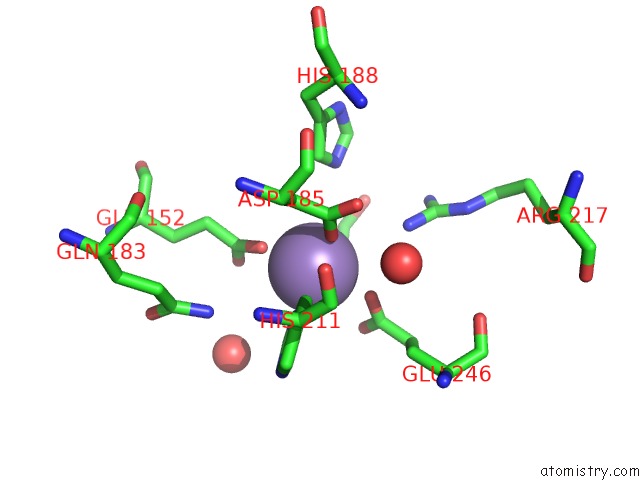
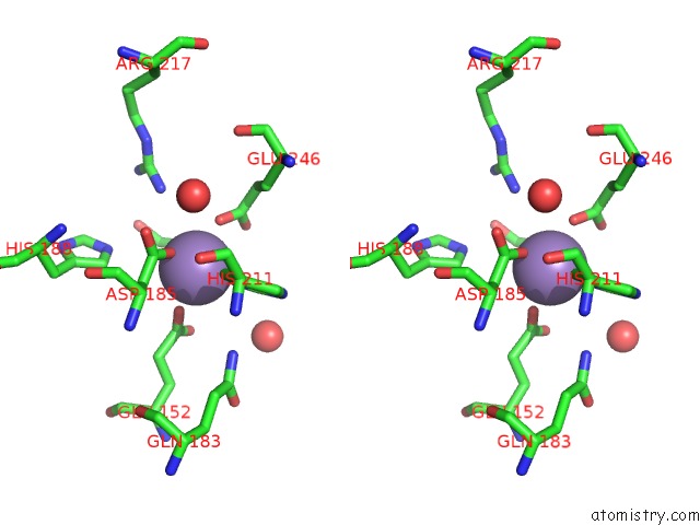
Manganese binding site 2 out of 4 in 4xsl
Go back to
Manganese binding site 2 out
of 4 in the Crystal Strcutre of D-Tagatose 3-Epimerase C66S From Pseudomonas Cichorii in Complex with Glycerol
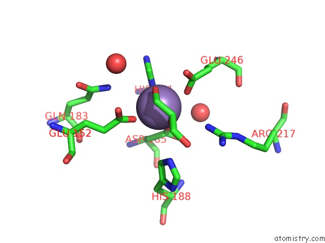
Mono view
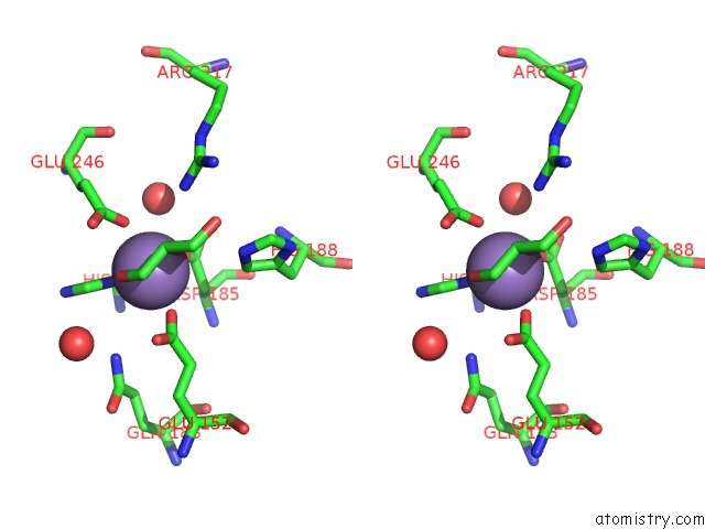
Stereo pair view
Mono view
Stereo pair view
A full contact list of Manganese with other atoms in the Mn binding
site number 2 of Crystal Strcutre of D-Tagatose 3-Epimerase C66S From Pseudomonas Cichorii in Complex with Glycerol within 5.0Å range:
|
Manganese binding site 3 out of 4 in 4xsl
Go back to
Manganese binding site 3 out
of 4 in the Crystal Strcutre of D-Tagatose 3-Epimerase C66S From Pseudomonas Cichorii in Complex with Glycerol
Mono view
Stereo pair view
Mono view
Stereo pair view
A full contact list of Manganese with other atoms in the Mn binding
site number 3 of Crystal Strcutre of D-Tagatose 3-Epimerase C66S From Pseudomonas Cichorii in Complex with Glycerol within 5.0Å range:
|
Manganese binding site 4 out of 4 in 4xsl
Go back to
Manganese binding site 4 out
of 4 in the Crystal Strcutre of D-Tagatose 3-Epimerase C66S From Pseudomonas Cichorii in Complex with Glycerol
Mono view
Stereo pair view
Mono view
Stereo pair view
A full contact list of Manganese with other atoms in the Mn binding
site number 4 of Crystal Strcutre of D-Tagatose 3-Epimerase C66S From Pseudomonas Cichorii in Complex with Glycerol within 5.0Å range:
|
Reference:
H.Yoshida,
A.Yoshihara,
T.Ishii,
K.Izumori,
S.Kamitori.
X-Ray Structures of the Pseudomonas Cichorii D-Tagatose 3-Epimerase Mutant Form C66S Recognizing Deoxy Sugars As Substrates Appl. Microbiol. Biotechnol. V. 100 10403 2016.
ISSN: ESSN 1432-0614
PubMed: 27368739
DOI: 10.1007/S00253-016-7673-7
Page generated: Sat Oct 5 22:56:10 2024
ISSN: ESSN 1432-0614
PubMed: 27368739
DOI: 10.1007/S00253-016-7673-7
Last articles
Zn in 9MJ5Zn in 9HNW
Zn in 9G0L
Zn in 9FNE
Zn in 9DZN
Zn in 9E0I
Zn in 9D32
Zn in 9DAK
Zn in 8ZXC
Zn in 8ZUF