Manganese »
PDB 4wl8-4xwt »
4xjk »
Manganese in PDB 4xjk: Crystal Structure of Mn(II) Ca(II) Na(I) Bound Calprotectin
Protein crystallography data
The structure of Crystal Structure of Mn(II) Ca(II) Na(I) Bound Calprotectin, PDB code: 4xjk
was solved by
C.L.Drennan,
S.E.J.Bowman,
with X-Ray Crystallography technique. A brief refinement statistics is given in the table below:
| Resolution Low / High (Å) | 49.24 / 1.76 |
| Space group | P 1 2 1 |
| Cell size a, b, c (Å), α, β, γ (°) | 55.279, 49.239, 218.067, 90.00, 94.07, 90.00 |
| R / Rfree (%) | 18.5 / 22 |
Other elements in 4xjk:
The structure of Crystal Structure of Mn(II) Ca(II) Na(I) Bound Calprotectin also contains other interesting chemical elements:
| Calcium | (Ca) | 10 atoms |
| Sodium | (Na) | 10 atoms |
Manganese Binding Sites:
The binding sites of Manganese atom in the Crystal Structure of Mn(II) Ca(II) Na(I) Bound Calprotectin
(pdb code 4xjk). This binding sites where shown within
5.0 Angstroms radius around Manganese atom.
In total 5 binding sites of Manganese where determined in the Crystal Structure of Mn(II) Ca(II) Na(I) Bound Calprotectin, PDB code: 4xjk:
Jump to Manganese binding site number: 1; 2; 3; 4; 5;
In total 5 binding sites of Manganese where determined in the Crystal Structure of Mn(II) Ca(II) Na(I) Bound Calprotectin, PDB code: 4xjk:
Jump to Manganese binding site number: 1; 2; 3; 4; 5;
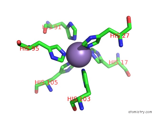
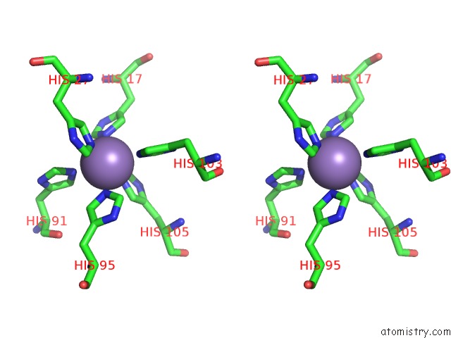
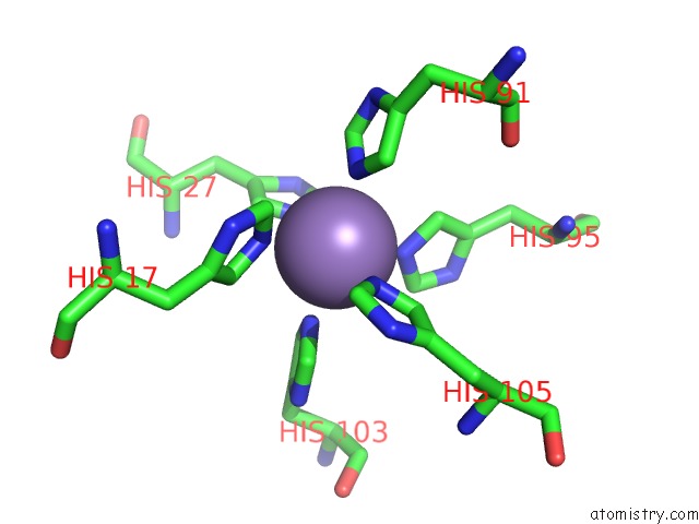
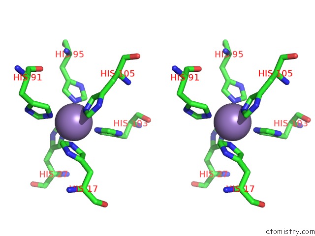
Manganese binding site 1 out of 5 in 4xjk
Go back to
Manganese binding site 1 out
of 5 in the Crystal Structure of Mn(II) Ca(II) Na(I) Bound Calprotectin
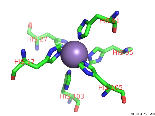
Mono view
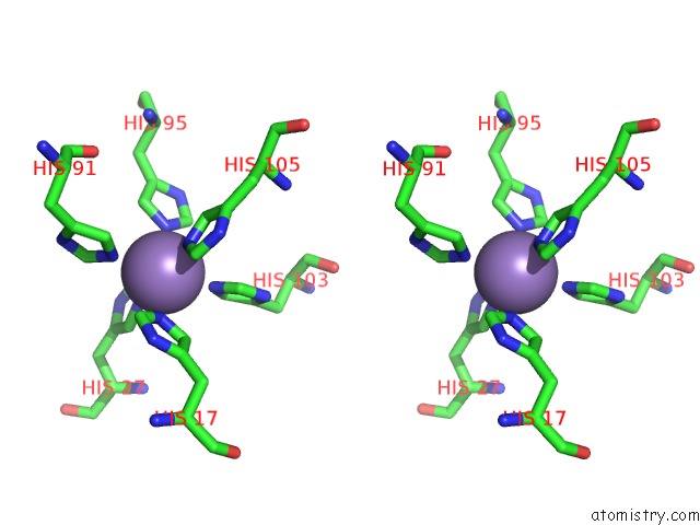
Stereo pair view
Mono view
Stereo pair view
A full contact list of Manganese with other atoms in the Mn binding
site number 1 of Crystal Structure of Mn(II) Ca(II) Na(I) Bound Calprotectin within 5.0Å range:
|
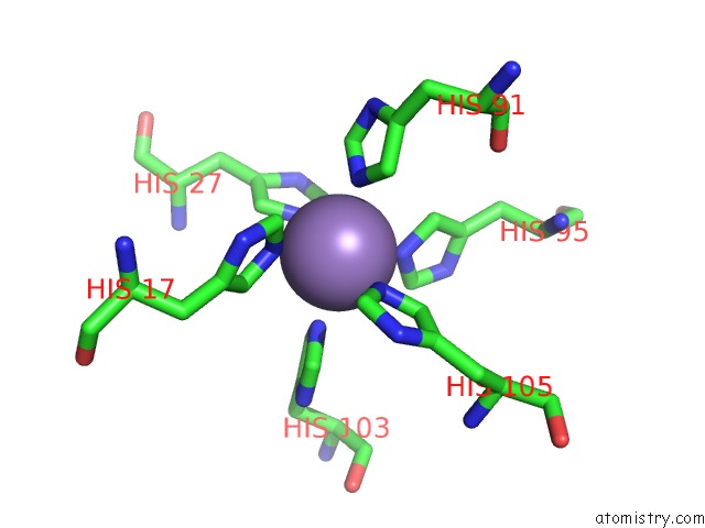
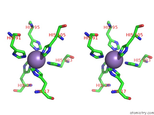
Manganese binding site 2 out of 5 in 4xjk
Go back to
Manganese binding site 2 out
of 5 in the Crystal Structure of Mn(II) Ca(II) Na(I) Bound Calprotectin
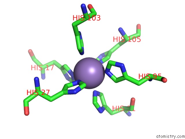
Mono view
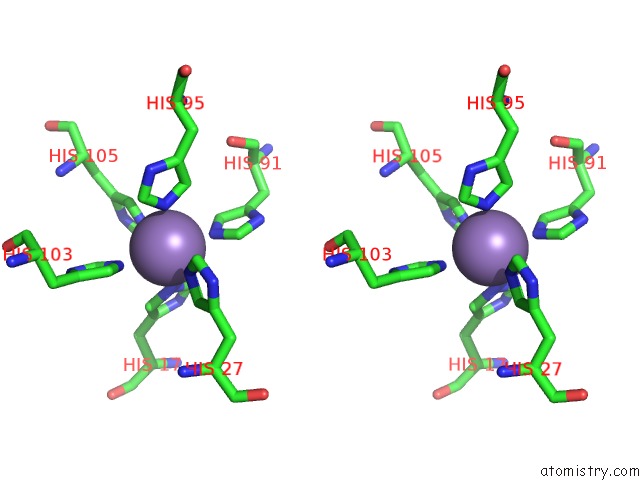
Stereo pair view
Mono view
Stereo pair view
A full contact list of Manganese with other atoms in the Mn binding
site number 2 of Crystal Structure of Mn(II) Ca(II) Na(I) Bound Calprotectin within 5.0Å range:
|
Manganese binding site 3 out of 5 in 4xjk
Go back to
Manganese binding site 3 out
of 5 in the Crystal Structure of Mn(II) Ca(II) Na(I) Bound Calprotectin
Mono view
Stereo pair view
Mono view
Stereo pair view
A full contact list of Manganese with other atoms in the Mn binding
site number 3 of Crystal Structure of Mn(II) Ca(II) Na(I) Bound Calprotectin within 5.0Å range:
|
Manganese binding site 4 out of 5 in 4xjk
Go back to
Manganese binding site 4 out
of 5 in the Crystal Structure of Mn(II) Ca(II) Na(I) Bound Calprotectin
Mono view
Stereo pair view
Mono view
Stereo pair view
A full contact list of Manganese with other atoms in the Mn binding
site number 4 of Crystal Structure of Mn(II) Ca(II) Na(I) Bound Calprotectin within 5.0Å range:
|
Manganese binding site 5 out of 5 in 4xjk
Go back to
Manganese binding site 5 out
of 5 in the Crystal Structure of Mn(II) Ca(II) Na(I) Bound Calprotectin
Mono view
Stereo pair view
Mono view
Stereo pair view
A full contact list of Manganese with other atoms in the Mn binding
site number 5 of Crystal Structure of Mn(II) Ca(II) Na(I) Bound Calprotectin within 5.0Å range:
|
Reference:
D.M.Gagnon,
M.B.Brophy,
S.E.Bowman,
T.A.Stich,
C.L.Drennan,
R.D.Britt,
E.M.Nolan.
Manganese Binding Properties of Human Calprotectin Under Conditions of High and Low Calcium: X-Ray Crystallographic and Advanced Electron Paramagnetic Resonance Spectroscopic Analysis. J.Am.Chem.Soc. V. 137 3004 2015.
ISSN: ESSN 1520-5126
PubMed: 25597447
DOI: 10.1021/JA512204S
Page generated: Sat Oct 5 22:55:06 2024
ISSN: ESSN 1520-5126
PubMed: 25597447
DOI: 10.1021/JA512204S
Last articles
Fe in 2YXOFe in 2YRS
Fe in 2YXC
Fe in 2YNM
Fe in 2YVJ
Fe in 2YP1
Fe in 2YU2
Fe in 2YU1
Fe in 2YQB
Fe in 2YOO