Manganese »
PDB 4wl8-4xwt »
4x9q »
Manganese in PDB 4x9q: Mnsod-3 Room Temperature Structure
Enzymatic activity of Mnsod-3 Room Temperature Structure
All present enzymatic activity of Mnsod-3 Room Temperature Structure:
1.15.1.1;
1.15.1.1;
Protein crystallography data
The structure of Mnsod-3 Room Temperature Structure, PDB code: 4x9q
was solved by
G.J.Hunter,
C.H.Trinh,
T.Hunter,
R.Bonetta,
E.E.Stewart,
with X-Ray Crystallography technique. A brief refinement statistics is given in the table below:
| Resolution Low / High (Å) | 70.19 / 1.77 |
| Space group | P 41 21 2 |
| Cell size a, b, c (Å), α, β, γ (°) | 81.465, 81.465, 137.959, 90.00, 90.00, 90.00 |
| R / Rfree (%) | 18.1 / 21.4 |
Manganese Binding Sites:
The binding sites of Manganese atom in the Mnsod-3 Room Temperature Structure
(pdb code 4x9q). This binding sites where shown within
5.0 Angstroms radius around Manganese atom.
In total 2 binding sites of Manganese where determined in the Mnsod-3 Room Temperature Structure, PDB code: 4x9q:
Jump to Manganese binding site number: 1; 2;
In total 2 binding sites of Manganese where determined in the Mnsod-3 Room Temperature Structure, PDB code: 4x9q:
Jump to Manganese binding site number: 1; 2;
Manganese binding site 1 out of 2 in 4x9q
Go back to
Manganese binding site 1 out
of 2 in the Mnsod-3 Room Temperature Structure
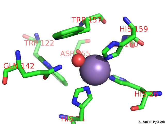
Mono view
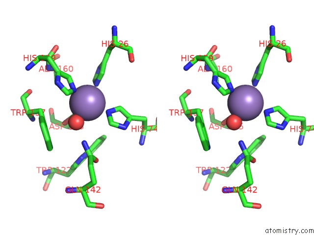
Stereo pair view
Mono view
Stereo pair view
A full contact list of Manganese with other atoms in the Mn binding
site number 1 of Mnsod-3 Room Temperature Structure within 5.0Å range:
|
Manganese binding site 2 out of 2 in 4x9q
Go back to
Manganese binding site 2 out
of 2 in the Mnsod-3 Room Temperature Structure
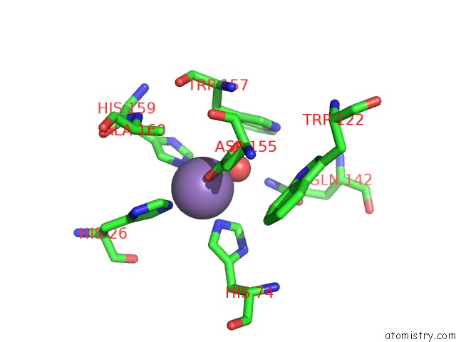
Mono view
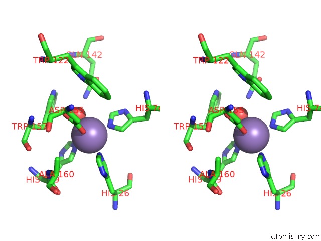
Stereo pair view
Mono view
Stereo pair view
A full contact list of Manganese with other atoms in the Mn binding
site number 2 of Mnsod-3 Room Temperature Structure within 5.0Å range:
|
Reference:
G.J.Hunter,
C.H.Trinh,
R.Bonetta,
E.E.Stewart,
D.E.Cabelli,
T.Hunter.
The Structure of the Caenorhabditis Elegans Manganese Superoxide Dismutase Mnsod-3-Azide Complex. Protein Sci. V. 24 1777 2015.
ISSN: ESSN 1469-896X
PubMed: 26257399
DOI: 10.1002/PRO.2768
Page generated: Sat Oct 5 22:54:36 2024
ISSN: ESSN 1469-896X
PubMed: 26257399
DOI: 10.1002/PRO.2768
Last articles
Zn in 9J0NZn in 9J0O
Zn in 9J0P
Zn in 9FJX
Zn in 9EKB
Zn in 9C0F
Zn in 9CAH
Zn in 9CH0
Zn in 9CH3
Zn in 9CH1