Manganese »
PDB 4wl8-4xwt »
4wlz »
Manganese in PDB 4wlz: Crystal Structure of Mouse Xyloside Xylosyltransferase 1 Complexed with Manganese and Udp
Protein crystallography data
The structure of Crystal Structure of Mouse Xyloside Xylosyltransferase 1 Complexed with Manganese and Udp, PDB code: 4wlz
was solved by
H.Yu,
H.Li,
with X-Ray Crystallography technique. A brief refinement statistics is given in the table below:
| Resolution Low / High (Å) | 50.00 / 3.03 |
| Space group | P 31 2 1 |
| Cell size a, b, c (Å), α, β, γ (°) | 89.662, 89.662, 154.987, 90.00, 90.00, 120.00 |
| R / Rfree (%) | 23 / 28.8 |
Manganese Binding Sites:
The binding sites of Manganese atom in the Crystal Structure of Mouse Xyloside Xylosyltransferase 1 Complexed with Manganese and Udp
(pdb code 4wlz). This binding sites where shown within
5.0 Angstroms radius around Manganese atom.
In total 2 binding sites of Manganese where determined in the Crystal Structure of Mouse Xyloside Xylosyltransferase 1 Complexed with Manganese and Udp, PDB code: 4wlz:
Jump to Manganese binding site number: 1; 2;
In total 2 binding sites of Manganese where determined in the Crystal Structure of Mouse Xyloside Xylosyltransferase 1 Complexed with Manganese and Udp, PDB code: 4wlz:
Jump to Manganese binding site number: 1; 2;
Manganese binding site 1 out of 2 in 4wlz
Go back to
Manganese binding site 1 out
of 2 in the Crystal Structure of Mouse Xyloside Xylosyltransferase 1 Complexed with Manganese and Udp
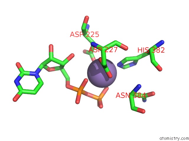
Mono view
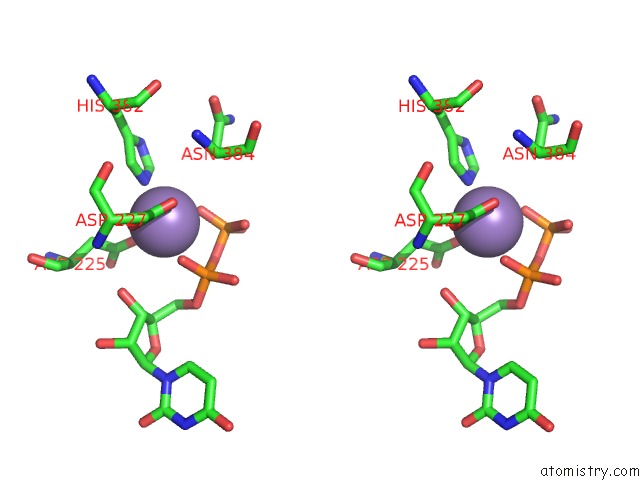
Stereo pair view
Mono view
Stereo pair view
A full contact list of Manganese with other atoms in the Mn binding
site number 1 of Crystal Structure of Mouse Xyloside Xylosyltransferase 1 Complexed with Manganese and Udp within 5.0Å range:
|
Manganese binding site 2 out of 2 in 4wlz
Go back to
Manganese binding site 2 out
of 2 in the Crystal Structure of Mouse Xyloside Xylosyltransferase 1 Complexed with Manganese and Udp
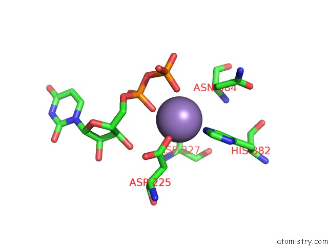
Mono view
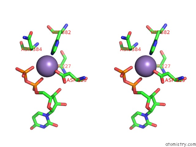
Stereo pair view
Mono view
Stereo pair view
A full contact list of Manganese with other atoms in the Mn binding
site number 2 of Crystal Structure of Mouse Xyloside Xylosyltransferase 1 Complexed with Manganese and Udp within 5.0Å range:
|
Reference:
H.Yu,
M.Takeuchi,
J.Lebarron,
J.Kantharia,
E.London,
H.Bakker,
R.S.Haltiwanger,
H.Li,
H.Takeuchi.
Notch-Modifying Xylosyltransferase Structures Support An Sni-Like Retaining Mechanism. Nat.Chem.Biol. V. 11 847 2015.
ISSN: ESSN 1552-4469
PubMed: 26414444
DOI: 10.1038/NCHEMBIO.1927
Page generated: Sat Oct 5 22:50:57 2024
ISSN: ESSN 1552-4469
PubMed: 26414444
DOI: 10.1038/NCHEMBIO.1927
Last articles
Cl in 3DJKCl in 3DJY
Cl in 3DIB
Cl in 3DIC
Cl in 3DI8
Cl in 3DI9
Cl in 3DI4
Cl in 3DI7
Cl in 3DI6
Cl in 3DHP