Manganese »
PDB 4u87-4wiu »
4u9n »
Manganese in PDB 4u9n: Structure of A Membrane Protein
Protein crystallography data
The structure of Structure of A Membrane Protein, PDB code: 4u9n
was solved by
H.Takeda,
M.Hattori,
T.Nishizawa,
K.Yamashita,
S.T.A.Shah,
M.Caffrey,
A.D.Maturana,
R.Ishitani,
O.Nureki,
with X-Ray Crystallography technique. A brief refinement statistics is given in the table below:
| Resolution Low / High (Å) | 41.71 / 2.20 |
| Space group | P 21 21 21 |
| Cell size a, b, c (Å), α, β, γ (°) | 63.521, 70.681, 103.327, 90.00, 90.00, 90.00 |
| R / Rfree (%) | 22.4 / 24 |
Manganese Binding Sites:
The binding sites of Manganese atom in the Structure of A Membrane Protein
(pdb code 4u9n). This binding sites where shown within
5.0 Angstroms radius around Manganese atom.
In total 4 binding sites of Manganese where determined in the Structure of A Membrane Protein, PDB code: 4u9n:
Jump to Manganese binding site number: 1; 2; 3; 4;
In total 4 binding sites of Manganese where determined in the Structure of A Membrane Protein, PDB code: 4u9n:
Jump to Manganese binding site number: 1; 2; 3; 4;
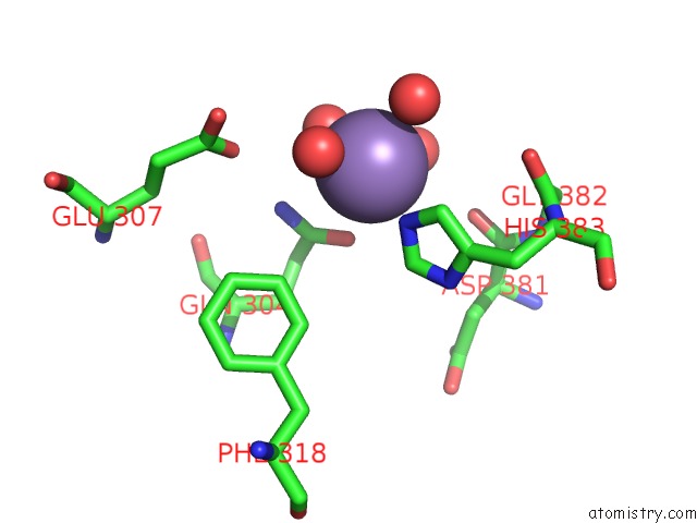
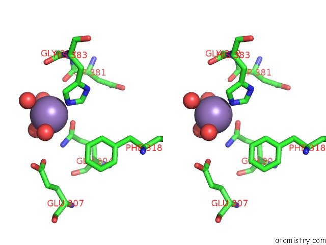
Manganese binding site 1 out of 4 in 4u9n
Go back to
Manganese binding site 1 out
of 4 in the Structure of A Membrane Protein
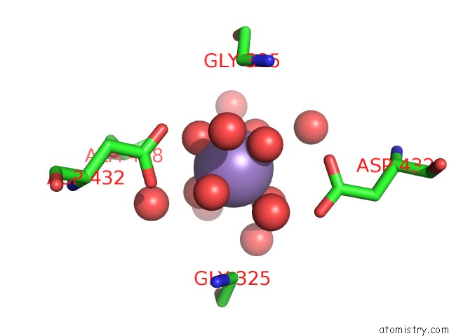
Mono view
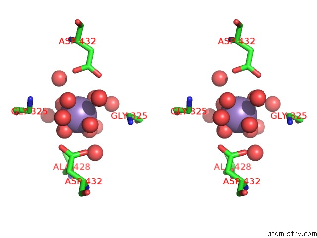
Stereo pair view
Mono view
Stereo pair view
A full contact list of Manganese with other atoms in the Mn binding
site number 1 of Structure of A Membrane Protein within 5.0Å range:
|
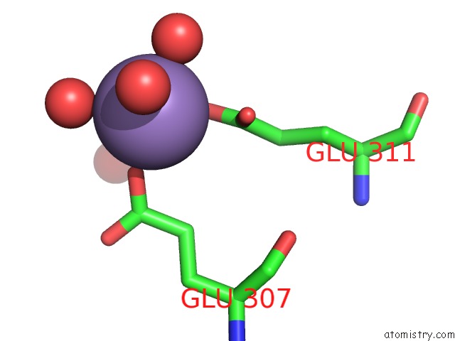
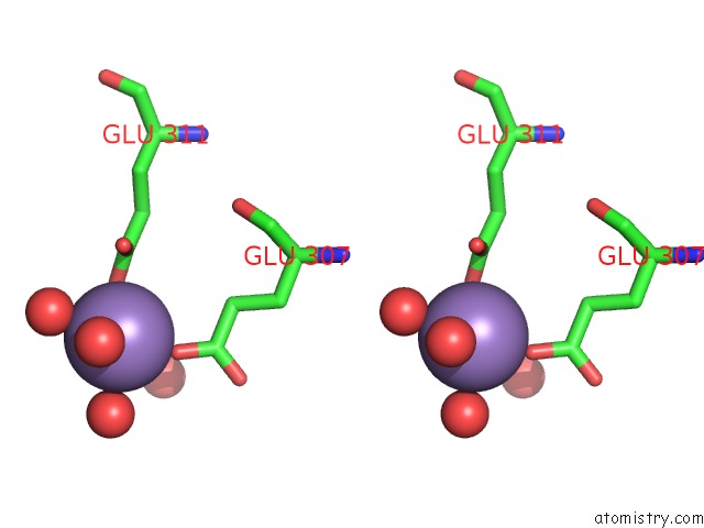
Manganese binding site 2 out of 4 in 4u9n
Go back to
Manganese binding site 2 out
of 4 in the Structure of A Membrane Protein
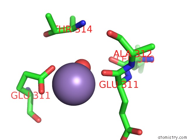
Mono view
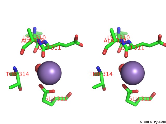
Stereo pair view
Mono view
Stereo pair view
A full contact list of Manganese with other atoms in the Mn binding
site number 2 of Structure of A Membrane Protein within 5.0Å range:
|
Manganese binding site 3 out of 4 in 4u9n
Go back to
Manganese binding site 3 out
of 4 in the Structure of A Membrane Protein
Mono view
Stereo pair view
Mono view
Stereo pair view
A full contact list of Manganese with other atoms in the Mn binding
site number 3 of Structure of A Membrane Protein within 5.0Å range:
|
Manganese binding site 4 out of 4 in 4u9n
Go back to
Manganese binding site 4 out
of 4 in the Structure of A Membrane Protein
Mono view
Stereo pair view
Mono view
Stereo pair view
A full contact list of Manganese with other atoms in the Mn binding
site number 4 of Structure of A Membrane Protein within 5.0Å range:
|
Reference:
H.Takeda,
M.Hattori,
T.Nishizawa,
K.Yamashita,
S.T.Shah,
M.Caffrey,
A.D.Maturana,
R.Ishitani,
O.Nureki.
Structural Basis For Ion Selectivity Revealed By High-Resolution Crystal Structure of Mg(2+) Channel Mgte Nat Commun V. 5 5374 2014.
ISSN: ESSN 2041-1723
PubMed: 25367295
DOI: 10.1038/NCOMMS6374
Page generated: Sat Oct 5 21:18:28 2024
ISSN: ESSN 2041-1723
PubMed: 25367295
DOI: 10.1038/NCOMMS6374
Last articles
Fe in 2YXOFe in 2YRS
Fe in 2YXC
Fe in 2YNM
Fe in 2YVJ
Fe in 2YP1
Fe in 2YU2
Fe in 2YU1
Fe in 2YQB
Fe in 2YOO