Manganese »
PDB 4qsh-4u3o »
4rhm »
Manganese in PDB 4rhm: Crystal Structure of T. Brucei Arginase-Like Protein Quadruple Mutant S149D/R151H/S153D/S226D
Enzymatic activity of Crystal Structure of T. Brucei Arginase-Like Protein Quadruple Mutant S149D/R151H/S153D/S226D
All present enzymatic activity of Crystal Structure of T. Brucei Arginase-Like Protein Quadruple Mutant S149D/R151H/S153D/S226D:
3.5.3.1;
3.5.3.1;
Protein crystallography data
The structure of Crystal Structure of T. Brucei Arginase-Like Protein Quadruple Mutant S149D/R151H/S153D/S226D, PDB code: 4rhm
was solved by
Y.Hai,
M.P.Barrett,
D.W.Christianson,
with X-Ray Crystallography technique. A brief refinement statistics is given in the table below:
| Resolution Low / High (Å) | 42.88 / 1.95 |
| Space group | C 1 2 1 |
| Cell size a, b, c (Å), α, β, γ (°) | 82.123, 137.350, 87.778, 90.00, 102.28, 90.00 |
| R / Rfree (%) | 20.1 / 23.7 |
Manganese Binding Sites:
The binding sites of Manganese atom in the Crystal Structure of T. Brucei Arginase-Like Protein Quadruple Mutant S149D/R151H/S153D/S226D
(pdb code 4rhm). This binding sites where shown within
5.0 Angstroms radius around Manganese atom.
In total 3 binding sites of Manganese where determined in the Crystal Structure of T. Brucei Arginase-Like Protein Quadruple Mutant S149D/R151H/S153D/S226D, PDB code: 4rhm:
Jump to Manganese binding site number: 1; 2; 3;
In total 3 binding sites of Manganese where determined in the Crystal Structure of T. Brucei Arginase-Like Protein Quadruple Mutant S149D/R151H/S153D/S226D, PDB code: 4rhm:
Jump to Manganese binding site number: 1; 2; 3;
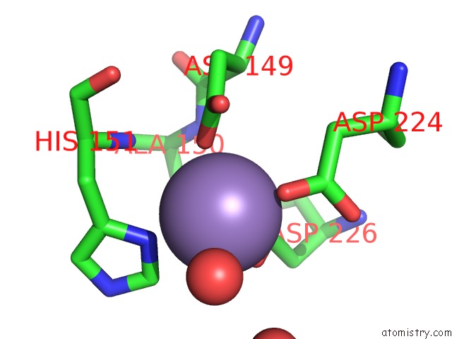
Manganese binding site 1 out of 3 in 4rhm
Go back to
Manganese binding site 1 out
of 3 in the Crystal Structure of T. Brucei Arginase-Like Protein Quadruple Mutant S149D/R151H/S153D/S226D
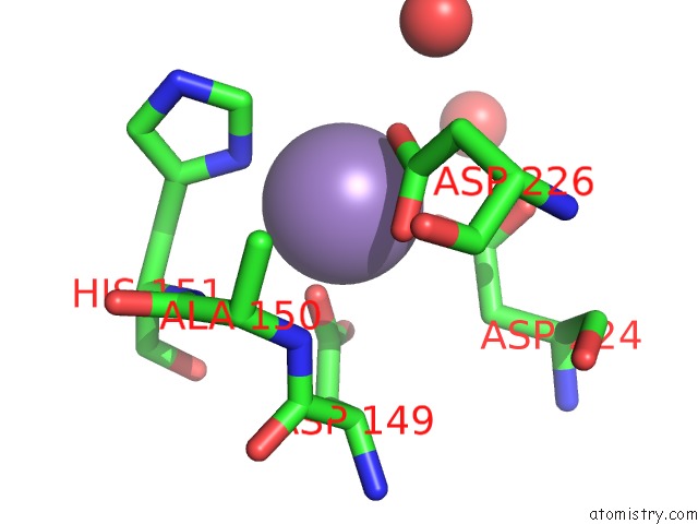
Mono view
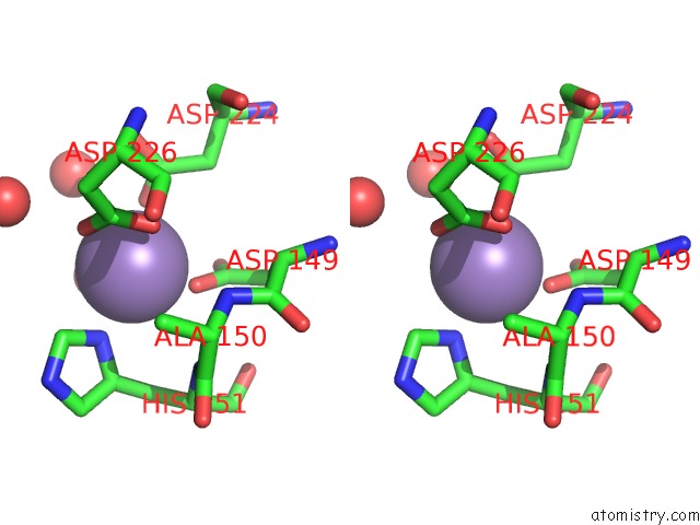
Stereo pair view
Mono view
Stereo pair view
A full contact list of Manganese with other atoms in the Mn binding
site number 1 of Crystal Structure of T. Brucei Arginase-Like Protein Quadruple Mutant S149D/R151H/S153D/S226D within 5.0Å range:
|
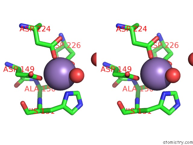
Manganese binding site 2 out of 3 in 4rhm
Go back to
Manganese binding site 2 out
of 3 in the Crystal Structure of T. Brucei Arginase-Like Protein Quadruple Mutant S149D/R151H/S153D/S226D
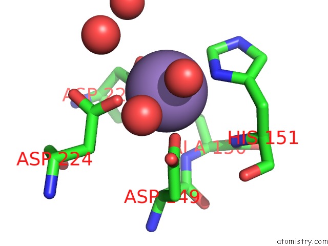
Mono view
Stereo pair view
Mono view
Stereo pair view
A full contact list of Manganese with other atoms in the Mn binding
site number 2 of Crystal Structure of T. Brucei Arginase-Like Protein Quadruple Mutant S149D/R151H/S153D/S226D within 5.0Å range:
|
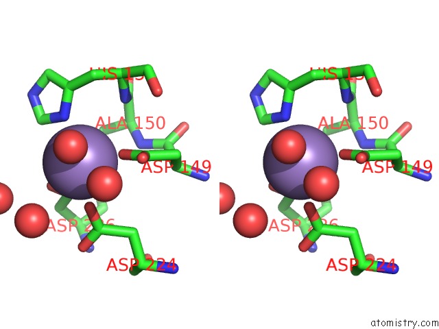
Manganese binding site 3 out of 3 in 4rhm
Go back to
Manganese binding site 3 out
of 3 in the Crystal Structure of T. Brucei Arginase-Like Protein Quadruple Mutant S149D/R151H/S153D/S226D
Mono view
Stereo pair view
Mono view
Stereo pair view
A full contact list of Manganese with other atoms in the Mn binding
site number 3 of Crystal Structure of T. Brucei Arginase-Like Protein Quadruple Mutant S149D/R151H/S153D/S226D within 5.0Å range:
|
Reference:
Y.Hai,
E.J.Kerkhoven,
M.P.Barrett,
D.W.Christianson.
Crystal Structure of An Arginase-Like Protein From Trypanosoma Brucei That Evolved Without A Binuclear Manganese Cluster. Biochemistry V. 54 458 2015.
ISSN: ISSN 0006-2960
PubMed: 25536859
DOI: 10.1021/BI501366A
Page generated: Sat Oct 5 21:11:14 2024
ISSN: ISSN 0006-2960
PubMed: 25536859
DOI: 10.1021/BI501366A
Last articles
Zn in 9J0NZn in 9J0O
Zn in 9J0P
Zn in 9FJX
Zn in 9EKB
Zn in 9C0F
Zn in 9CAH
Zn in 9CH0
Zn in 9CH3
Zn in 9CH1