Manganese »
PDB 4php-4qsf »
4q42 »
Manganese in PDB 4q42: Crystal Structure of Schistosoma Mansoni Arginase in Complex with L- Ornithine
Enzymatic activity of Crystal Structure of Schistosoma Mansoni Arginase in Complex with L- Ornithine
All present enzymatic activity of Crystal Structure of Schistosoma Mansoni Arginase in Complex with L- Ornithine:
3.5.3.1;
3.5.3.1;
Protein crystallography data
The structure of Crystal Structure of Schistosoma Mansoni Arginase in Complex with L- Ornithine, PDB code: 4q42
was solved by
Y.Hai,
J.E.Edwards,
M.C.Van Zandt,
K.F.Hoffmann,
D.W.Christianson,
with X-Ray Crystallography technique. A brief refinement statistics is given in the table below:
| Resolution Low / High (Å) | 49.49 / 2.05 |
| Space group | P 21 3 |
| Cell size a, b, c (Å), α, β, γ (°) | 178.447, 178.447, 178.447, 90.00, 90.00, 90.00 |
| R / Rfree (%) | 17.4 / 20.8 |
Manganese Binding Sites:
The binding sites of Manganese atom in the Crystal Structure of Schistosoma Mansoni Arginase in Complex with L- Ornithine
(pdb code 4q42). This binding sites where shown within
5.0 Angstroms radius around Manganese atom.
In total 8 binding sites of Manganese where determined in the Crystal Structure of Schistosoma Mansoni Arginase in Complex with L- Ornithine, PDB code: 4q42:
Jump to Manganese binding site number: 1; 2; 3; 4; 5; 6; 7; 8;
In total 8 binding sites of Manganese where determined in the Crystal Structure of Schistosoma Mansoni Arginase in Complex with L- Ornithine, PDB code: 4q42:
Jump to Manganese binding site number: 1; 2; 3; 4; 5; 6; 7; 8;
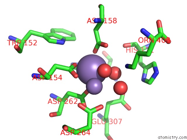
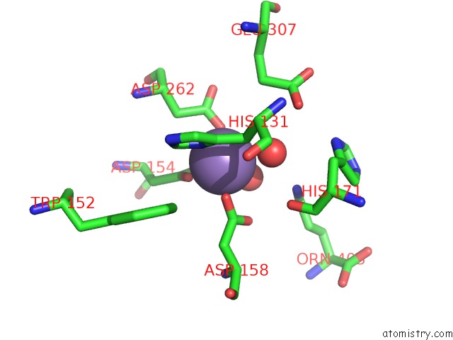
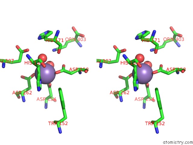
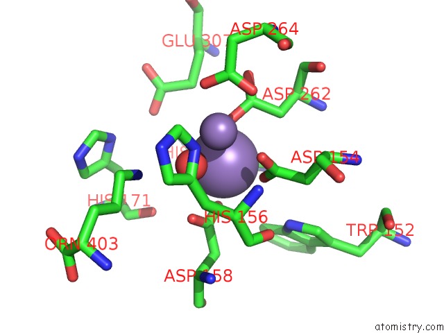
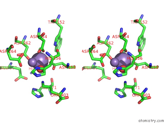
Manganese binding site 1 out of 8 in 4q42
Go back to
Manganese binding site 1 out
of 8 in the Crystal Structure of Schistosoma Mansoni Arginase in Complex with L- Ornithine
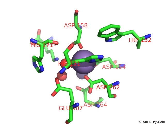
Mono view
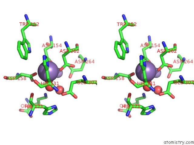
Stereo pair view
Mono view
Stereo pair view
A full contact list of Manganese with other atoms in the Mn binding
site number 1 of Crystal Structure of Schistosoma Mansoni Arginase in Complex with L- Ornithine within 5.0Å range:
|
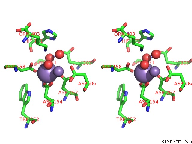
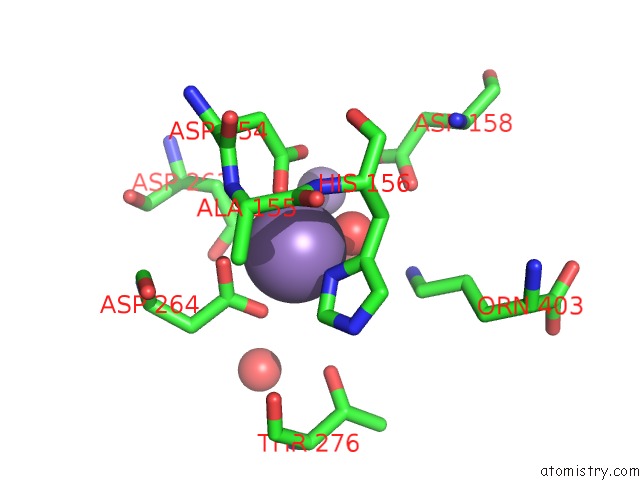
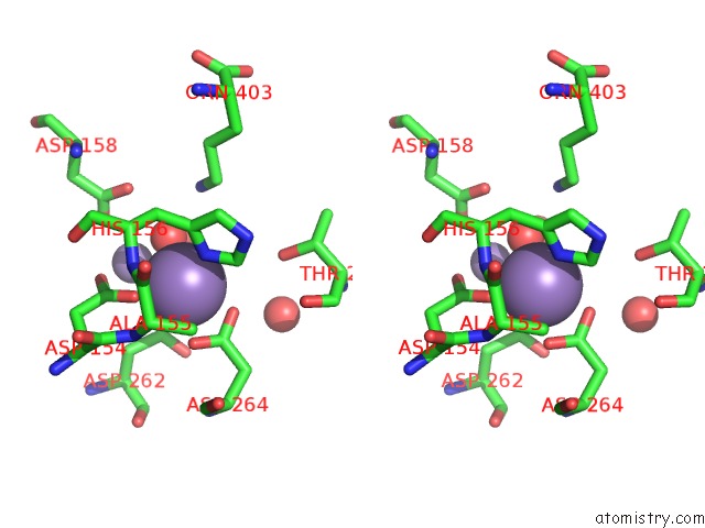
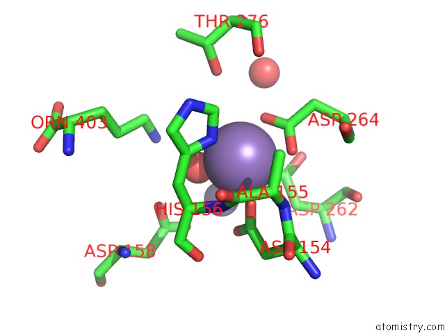
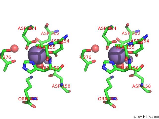
Manganese binding site 2 out of 8 in 4q42
Go back to
Manganese binding site 2 out
of 8 in the Crystal Structure of Schistosoma Mansoni Arginase in Complex with L- Ornithine
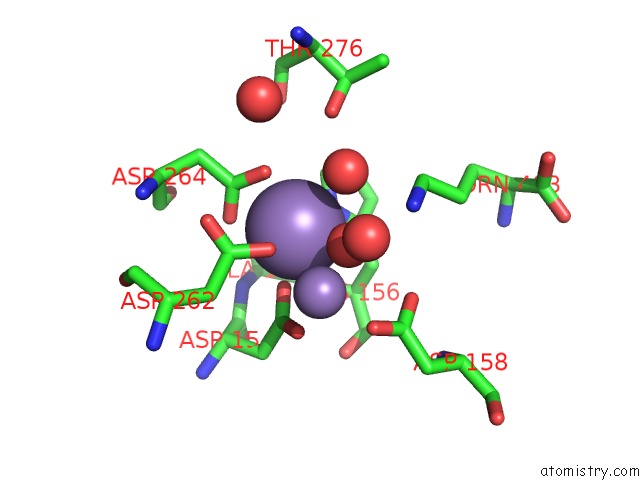
Mono view
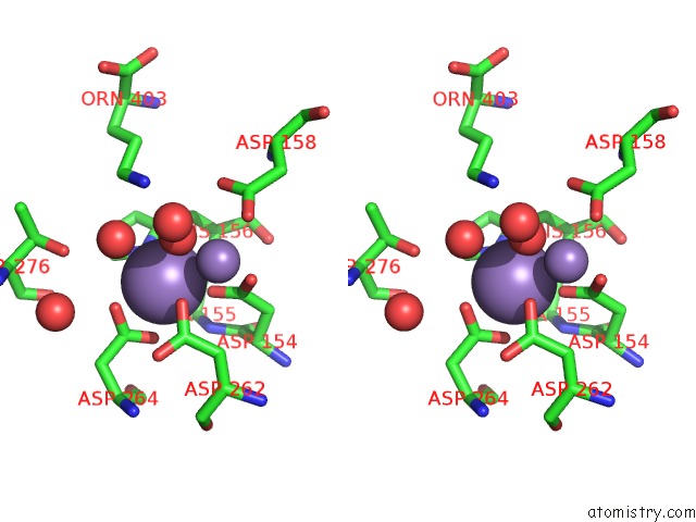
Stereo pair view
Mono view
Stereo pair view
A full contact list of Manganese with other atoms in the Mn binding
site number 2 of Crystal Structure of Schistosoma Mansoni Arginase in Complex with L- Ornithine within 5.0Å range:
|
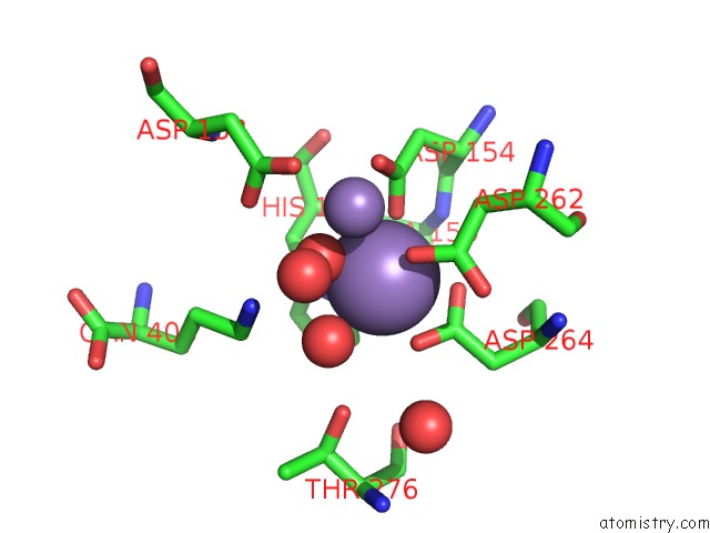
Manganese binding site 3 out of 8 in 4q42
Go back to
Manganese binding site 3 out
of 8 in the Crystal Structure of Schistosoma Mansoni Arginase in Complex with L- Ornithine
Mono view
Stereo pair view
Mono view
Stereo pair view
A full contact list of Manganese with other atoms in the Mn binding
site number 3 of Crystal Structure of Schistosoma Mansoni Arginase in Complex with L- Ornithine within 5.0Å range:
|
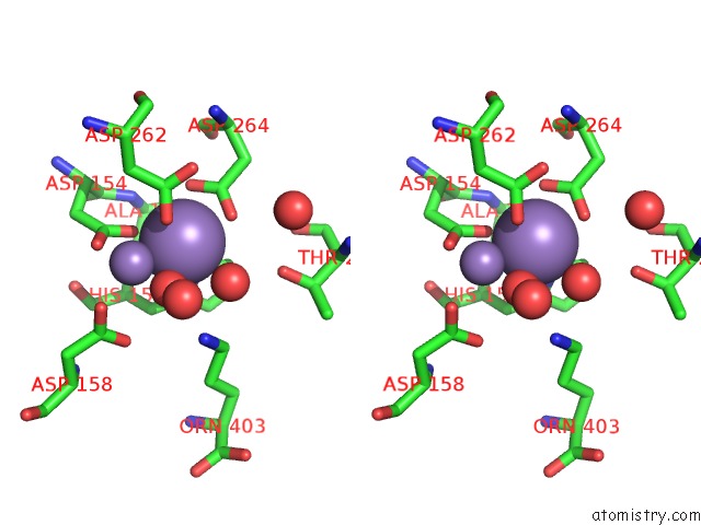
Manganese binding site 4 out of 8 in 4q42
Go back to
Manganese binding site 4 out
of 8 in the Crystal Structure of Schistosoma Mansoni Arginase in Complex with L- Ornithine
Mono view
Stereo pair view
Mono view
Stereo pair view
A full contact list of Manganese with other atoms in the Mn binding
site number 4 of Crystal Structure of Schistosoma Mansoni Arginase in Complex with L- Ornithine within 5.0Å range:
|
Manganese binding site 5 out of 8 in 4q42
Go back to
Manganese binding site 5 out
of 8 in the Crystal Structure of Schistosoma Mansoni Arginase in Complex with L- Ornithine
Mono view
Stereo pair view
Mono view
Stereo pair view
A full contact list of Manganese with other atoms in the Mn binding
site number 5 of Crystal Structure of Schistosoma Mansoni Arginase in Complex with L- Ornithine within 5.0Å range:
|
Manganese binding site 6 out of 8 in 4q42
Go back to
Manganese binding site 6 out
of 8 in the Crystal Structure of Schistosoma Mansoni Arginase in Complex with L- Ornithine
Mono view
Stereo pair view
Mono view
Stereo pair view
A full contact list of Manganese with other atoms in the Mn binding
site number 6 of Crystal Structure of Schistosoma Mansoni Arginase in Complex with L- Ornithine within 5.0Å range:
|
Manganese binding site 7 out of 8 in 4q42
Go back to
Manganese binding site 7 out
of 8 in the Crystal Structure of Schistosoma Mansoni Arginase in Complex with L- Ornithine
Mono view
Stereo pair view
Mono view
Stereo pair view
A full contact list of Manganese with other atoms in the Mn binding
site number 7 of Crystal Structure of Schistosoma Mansoni Arginase in Complex with L- Ornithine within 5.0Å range:
|
Manganese binding site 8 out of 8 in 4q42
Go back to
Manganese binding site 8 out
of 8 in the Crystal Structure of Schistosoma Mansoni Arginase in Complex with L- Ornithine
Mono view
Stereo pair view
Mono view
Stereo pair view
A full contact list of Manganese with other atoms in the Mn binding
site number 8 of Crystal Structure of Schistosoma Mansoni Arginase in Complex with L- Ornithine within 5.0Å range:
|
Reference:
Y.Hai,
J.E.Edwards,
M.C.Van Zandt,
K.F.Hoffmann,
D.W.Christianson.
Crystal Structure of Schistosoma Mansoni Arginase, A Potential Drug Target For the Treatment of Schistosomiasis. Biochemistry V. 53 4671 2014.
ISSN: ISSN 0006-2960
PubMed: 25007099
DOI: 10.1021/BI5004519
Page generated: Sat Oct 5 20:57:20 2024
ISSN: ISSN 0006-2960
PubMed: 25007099
DOI: 10.1021/BI5004519
Last articles
Zn in 9J0NZn in 9J0O
Zn in 9J0P
Zn in 9FJX
Zn in 9EKB
Zn in 9C0F
Zn in 9CAH
Zn in 9CH0
Zn in 9CH3
Zn in 9CH1