Manganese in PDB 4ptj: Ensemble Model For Escherichia Coli Dihydrofolate Reductase at 277K
Enzymatic activity of Ensemble Model For Escherichia Coli Dihydrofolate Reductase at 277K
All present enzymatic activity of Ensemble Model For Escherichia Coli Dihydrofolate Reductase at 277K:
1.5.1.3;
Protein crystallography data
The structure of Ensemble Model For Escherichia Coli Dihydrofolate Reductase at 277K, PDB code: 4ptj
was solved by
D.A.Keedy,
H.Van Den Bedem,
D.A.Sivak,
G.A.Petsko,
D.Ringe,
M.A.Wilson,
J.S.Fraser,
with X-Ray Crystallography technique. A brief refinement statistics is given in the table below:
Resolution Low / High (Å)
|
49.40 /
1.05
|
Space group
|
P 21 21 21
|
Cell size a, b, c (Å), α, β, γ (°)
|
34.299,
45.521,
98.711,
90.00,
90.00,
90.00
|
R / Rfree (%)
|
13.6 /
16.6
|
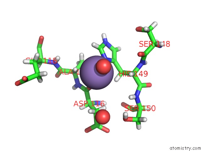
Manganese Binding Sites:
The binding sites of Manganese atom in the Ensemble Model For Escherichia Coli Dihydrofolate Reductase at 277K
(pdb code 4ptj). This binding sites where shown within
5.0 Angstroms radius around Manganese atom.
In total 2 binding sites of Manganese where determined in the
Ensemble Model For Escherichia Coli Dihydrofolate Reductase at 277K, PDB code: 4ptj:
Jump to Manganese binding site number:
1;
2;
Manganese binding site 1 out
of 2 in 4ptj
Go back to
Manganese Binding Sites List in 4ptj
Manganese binding site 1 out
of 2 in the Ensemble Model For Escherichia Coli Dihydrofolate Reductase at 277K
 Mono view
 Stereo pair view
|
A full contact list of Manganese with other atoms in the Mn binding
site number 1 of Ensemble Model For Escherichia Coli Dihydrofolate Reductase at 277K within 5.0Å range:
probe
|
atom
|
residue
|
distance (Å)
|
B
|
Occ
|
A:Mn203
b:14.0
occ:0.01
|
O
|
A:HOH366
|
2.2
|
14.8
|
0.0
|
O
|
A:ASP116
|
2.2
|
13.1
|
0.0
|
ND1
|
A:HIS149
|
2.3
|
13.5
|
0.0
|
CE1
|
A:HIS149
|
3.1
|
13.7
|
0.0
|
HE1
|
A:HIS149
|
3.1
|
13.9
|
0.0
|
HA
|
A:ALA117
|
3.2
|
13.1
|
0.0
|
C
|
A:ASP116
|
3.4
|
12.7
|
0.0
|
CG
|
A:HIS149
|
3.4
|
13.1
|
0.0
|
HB3
|
A:HIS149
|
3.4
|
12.9
|
0.0
|
HA
|
A:HIS149
|
3.7
|
13.8
|
0.0
|
CB
|
A:HIS149
|
3.8
|
12.9
|
0.0
|
CA
|
A:ALA117
|
3.9
|
12.7
|
0.0
|
N
|
A:ALA117
|
4.1
|
12.5
|
0.0
|
HB2
|
A:ALA117
|
4.2
|
12.4
|
0.0
|
HB2
|
A:ASP116
|
4.2
|
13.0
|
0.0
|
HB3
|
A:ASP116
|
4.2
|
13.2
|
0.0
|
CA
|
A:HIS149
|
4.3
|
13.5
|
0.0
|
NE2
|
A:HIS149
|
4.3
|
13.3
|
0.0
|
HB1
|
A:ALA117
|
4.4
|
12.7
|
0.0
|
CB
|
A:ALA117
|
4.4
|
12.5
|
0.0
|
CD2
|
A:HIS149
|
4.5
|
13.0
|
0.0
|
H
|
A:SER150
|
4.5
|
13.6
|
0.0
|
O
|
A:HOH328
|
4.5
|
14.2
|
0.0
|
CB
|
A:ASP116
|
4.5
|
12.9
|
0.0
|
CA
|
A:ASP116
|
4.6
|
12.5
|
0.0
|
HB2
|
A:HIS149
|
4.7
|
12.6
|
0.0
|
O
|
A:SER148
|
4.8
|
14.6
|
0.0
|
H
|
A:GLU118
|
4.8
|
13.6
|
0.0
|
H
|
A:ALA117
|
4.9
|
12.3
|
0.0
|
|
Manganese binding site 2 out
of 2 in 4ptj
Go back to
Manganese Binding Sites List in 4ptj
Manganese binding site 2 out
of 2 in the Ensemble Model For Escherichia Coli Dihydrofolate Reductase at 277K
 Mono view
 Stereo pair view
|
A full contact list of Manganese with other atoms in the Mn binding
site number 2 of Ensemble Model For Escherichia Coli Dihydrofolate Reductase at 277K within 5.0Å range:
probe
|
atom
|
residue
|
distance (Å)
|
B
|
Occ
|
A:Mn204
b:10.4
occ:0.01
|
HG
|
A:LEU156
|
1.6
|
10.9
|
0.0
|
OE2
|
A:GLU154
|
2.1
|
11.1
|
0.0
|
CG
|
A:LEU156
|
2.5
|
11.1
|
0.0
|
HD13
|
A:LEU112
|
2.6
|
10.2
|
0.0
|
CD
|
A:GLU154
|
2.6
|
11.2
|
0.0
|
OE1
|
A:GLU154
|
2.7
|
11.2
|
0.0
|
HG
|
A:LEU112
|
2.8
|
9.7
|
0.0
|
HD13
|
A:LEU156
|
2.8
|
10.6
|
0.0
|
HB2
|
A:LEU110
|
2.9
|
10.1
|
0.0
|
H
|
A:LEU156
|
2.9
|
10.8
|
0.0
|
HB2
|
A:LEU156
|
2.9
|
10.9
|
0.0
|
CD1
|
A:LEU156
|
2.9
|
10.8
|
0.0
|
HD11
|
A:LEU156
|
3.0
|
10.8
|
0.0
|
O
|
A:LEU110
|
3.1
|
10.2
|
0.0
|
CB
|
A:LEU156
|
3.1
|
11.2
|
0.0
|
HB2
|
A:LEU112
|
3.2
|
10.0
|
0.0
|
N
|
A:LEU156
|
3.2
|
11.1
|
0.0
|
CD1
|
A:LEU112
|
3.4
|
10.0
|
0.0
|
CG
|
A:LEU112
|
3.4
|
9.7
|
0.0
|
O
|
A:GLU154
|
3.4
|
10.4
|
0.0
|
HB3
|
A:GLU154
|
3.5
|
10.8
|
0.0
|
H
|
A:LEU112
|
3.5
|
9.9
|
0.0
|
HA
|
A:ILE155
|
3.6
|
10.7
|
0.0
|
CD2
|
A:LEU156
|
3.7
|
11.6
|
0.0
|
CB
|
A:LEU110
|
3.7
|
9.9
|
0.0
|
CA
|
A:LEU156
|
3.7
|
11.4
|
0.0
|
HB3
|
A:LEU110
|
3.7
|
9.7
|
0.0
|
C
|
A:ILE155
|
3.8
|
11.3
|
0.0
|
CB
|
A:LEU112
|
3.8
|
9.8
|
0.0
|
HD22
|
A:LEU156
|
3.8
|
11.8
|
0.0
|
C
|
A:GLU154
|
3.8
|
10.8
|
0.0
|
C
|
A:LEU110
|
3.9
|
9.9
|
0.0
|
HD12
|
A:LEU156
|
3.9
|
11.0
|
0.0
|
CG
|
A:GLU154
|
3.9
|
11.4
|
0.0
|
HD12
|
A:LEU112
|
3.9
|
9.9
|
0.0
|
HD23
|
A:LEU156
|
4.0
|
11.6
|
0.0
|
HB3
|
A:LEU156
|
4.0
|
11.4
|
0.0
|
CA
|
A:ILE155
|
4.0
|
11.0
|
0.0
|
N
|
A:LEU112
|
4.0
|
9.7
|
0.0
|
HD11
|
A:LEU112
|
4.0
|
10.0
|
0.0
|
CB
|
A:GLU154
|
4.1
|
11.1
|
0.0
|
HD12
|
A:LEU110
|
4.1
|
10.1
|
0.0
|
N
|
A:ILE155
|
4.1
|
11.1
|
0.0
|
HA
|
A:LEU156
|
4.3
|
11.7
|
0.0
|
HG2
|
A:GLU154
|
4.3
|
11.7
|
0.0
|
HA
|
A:TYR111
|
4.4
|
9.9
|
0.0
|
CA
|
A:LEU110
|
4.4
|
9.8
|
0.0
|
HD21
|
A:LEU156
|
4.5
|
11.8
|
0.0
|
CA
|
A:LEU112
|
4.5
|
9.7
|
0.0
|
HD11
|
A:LEU110
|
4.5
|
10.4
|
0.0
|
HG3
|
A:GLU154
|
4.5
|
11.6
|
0.0
|
O
|
A:ILE155
|
4.5
|
11.8
|
0.0
|
HB3
|
A:LEU112
|
4.6
|
9.8
|
0.0
|
CA
|
A:GLU154
|
4.6
|
11.1
|
0.0
|
N
|
A:TYR111
|
4.6
|
9.6
|
0.0
|
CD1
|
A:LEU110
|
4.7
|
10.2
|
0.0
|
H
|
A:LEU110
|
4.7
|
10.4
|
0.0
|
H
|
A:ILE155
|
4.8
|
11.4
|
0.0
|
CD2
|
A:LEU112
|
4.8
|
9.5
|
0.0
|
CG
|
A:LEU110
|
4.8
|
10.0
|
0.0
|
C
|
A:TYR111
|
4.8
|
9.6
|
0.0
|
HD21
|
A:LEU112
|
4.8
|
9.4
|
0.0
|
CA
|
A:TYR111
|
4.8
|
9.7
|
0.0
|
C
|
A:LEU156
|
4.9
|
11.4
|
0.0
|
HB2
|
A:GLU154
|
4.9
|
11.3
|
0.0
|
O
|
A:LEU156
|
5.0
|
11.0
|
0.0
|
|
Reference:
D.A.Keedy,
H.Van Den Bedem,
D.A.Sivak,
G.A.Petsko,
D.Ringe,
M.A.Wilson,
J.S.Fraser.
Crystal Cryocooling Distorts Conformational Heterogeneity in A Model Michaelis Complex of Dhfr. Structure V. 22 899 2014.
ISSN: ISSN 0969-2126
PubMed: 24882744
DOI: 10.1016/J.STR.2014.04.016
Page generated: Sat Oct 5 20:52:08 2024
|