Manganese »
PDB 4nxz-4phd »
4ogc »
Manganese in PDB 4ogc: Crystal Structure of the Type II-C CAS9 Enzyme From Actinomyces Naeslundii
Protein crystallography data
The structure of Crystal Structure of the Type II-C CAS9 Enzyme From Actinomyces Naeslundii, PDB code: 4ogc
was solved by
F.Jiang,
E.Ma,
S.Lin,
J.A.Doudna,
with X-Ray Crystallography technique. A brief refinement statistics is given in the table below:
| Resolution Low / High (Å) | 68.30 / 2.80 |
| Space group | P 1 21 1 |
| Cell size a, b, c (Å), α, β, γ (°) | 74.610, 132.560, 80.040, 90.00, 95.38, 90.00 |
| R / Rfree (%) | 19.4 / 23.3 |
Other elements in 4ogc:
The structure of Crystal Structure of the Type II-C CAS9 Enzyme From Actinomyces Naeslundii also contains other interesting chemical elements:
| Magnesium | (Mg) | 2 atoms |
| Zinc | (Zn) | 1 atom |
Manganese Binding Sites:
The binding sites of Manganese atom in the Crystal Structure of the Type II-C CAS9 Enzyme From Actinomyces Naeslundii
(pdb code 4ogc). This binding sites where shown within
5.0 Angstroms radius around Manganese atom.
In total 2 binding sites of Manganese where determined in the Crystal Structure of the Type II-C CAS9 Enzyme From Actinomyces Naeslundii, PDB code: 4ogc:
Jump to Manganese binding site number: 1; 2;
In total 2 binding sites of Manganese where determined in the Crystal Structure of the Type II-C CAS9 Enzyme From Actinomyces Naeslundii, PDB code: 4ogc:
Jump to Manganese binding site number: 1; 2;
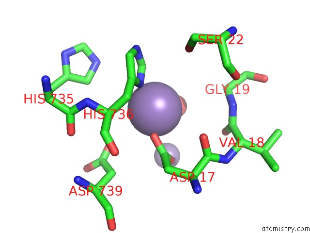
Manganese binding site 1 out of 2 in 4ogc
Go back to
Manganese binding site 1 out
of 2 in the Crystal Structure of the Type II-C CAS9 Enzyme From Actinomyces Naeslundii
Mono view
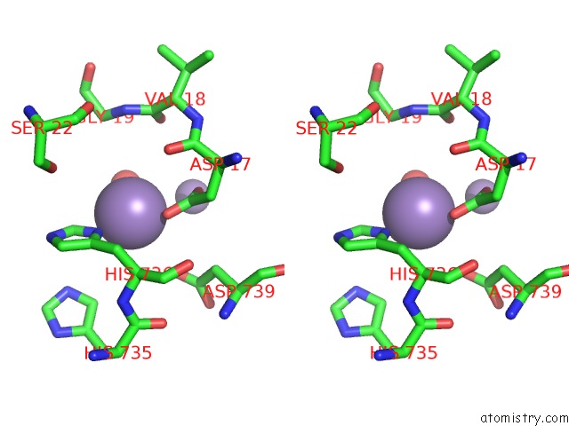
Stereo pair view
Mono view
Stereo pair view
A full contact list of Manganese with other atoms in the Mn binding
site number 1 of Crystal Structure of the Type II-C CAS9 Enzyme From Actinomyces Naeslundii within 5.0Å range:
|
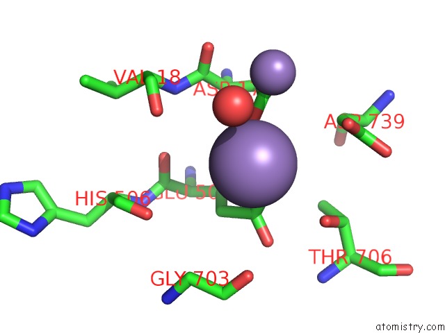
Manganese binding site 2 out of 2 in 4ogc
Go back to
Manganese binding site 2 out
of 2 in the Crystal Structure of the Type II-C CAS9 Enzyme From Actinomyces Naeslundii
Mono view
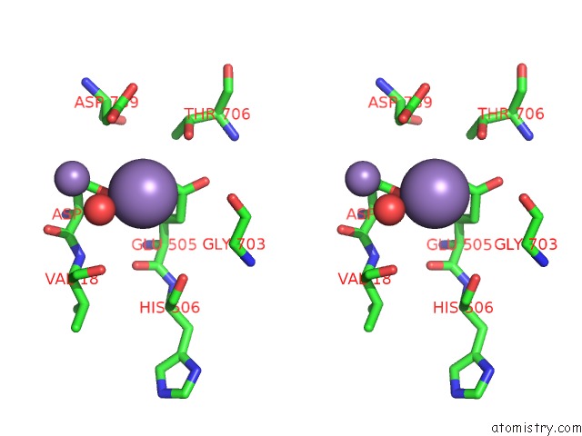
Stereo pair view
Mono view
Stereo pair view
A full contact list of Manganese with other atoms in the Mn binding
site number 2 of Crystal Structure of the Type II-C CAS9 Enzyme From Actinomyces Naeslundii within 5.0Å range:
|
Reference:
M.Jinek,
F.Jiang,
D.W.Taylor,
S.H.Sternberg,
E.Kaya,
E.Ma,
C.Anders,
M.Hauer,
K.Zhou,
S.Lin,
M.Kaplan,
A.T.Iavarone,
E.Charpentier,
E.Nogales,
J.A.Doudna.
Structures of CAS9 Endonucleases Reveal Rna-Mediated Conformational Activation. Science V. 343 47997 2014.
ISSN: ISSN 0036-8075
PubMed: 24505130
DOI: 10.1126/SCIENCE.1247997
Page generated: Sat Oct 5 20:42:16 2024
ISSN: ISSN 0036-8075
PubMed: 24505130
DOI: 10.1126/SCIENCE.1247997
Last articles
Zn in 9J0NZn in 9J0O
Zn in 9J0P
Zn in 9FJX
Zn in 9EKB
Zn in 9C0F
Zn in 9CAH
Zn in 9CH0
Zn in 9CH3
Zn in 9CH1