Manganese »
PDB 4mu4-4nx7 »
4nwx »
Manganese in PDB 4nwx: Crystal Structure of Phosphoglycerate Mutase From Staphylococcus Aureus in 2-Phosphoglyceric Acid Bound Form
Enzymatic activity of Crystal Structure of Phosphoglycerate Mutase From Staphylococcus Aureus in 2-Phosphoglyceric Acid Bound Form
All present enzymatic activity of Crystal Structure of Phosphoglycerate Mutase From Staphylococcus Aureus in 2-Phosphoglyceric Acid Bound Form:
5.4.2.12;
5.4.2.12;
Protein crystallography data
The structure of Crystal Structure of Phosphoglycerate Mutase From Staphylococcus Aureus in 2-Phosphoglyceric Acid Bound Form, PDB code: 4nwx
was solved by
A.Roychowdhury,
A.Kundu,
M.Bose,
A.Gujar,
A.K.Das,
with X-Ray Crystallography technique. A brief refinement statistics is given in the table below:
| Resolution Low / High (Å) | 19.64 / 2.01 |
| Space group | P 21 21 21 |
| Cell size a, b, c (Å), α, β, γ (°) | 73.639, 83.206, 89.073, 90.00, 90.00, 90.00 |
| R / Rfree (%) | 16.8 / 21.6 |
Manganese Binding Sites:
The binding sites of Manganese atom in the Crystal Structure of Phosphoglycerate Mutase From Staphylococcus Aureus in 2-Phosphoglyceric Acid Bound Form
(pdb code 4nwx). This binding sites where shown within
5.0 Angstroms radius around Manganese atom.
In total 2 binding sites of Manganese where determined in the Crystal Structure of Phosphoglycerate Mutase From Staphylococcus Aureus in 2-Phosphoglyceric Acid Bound Form, PDB code: 4nwx:
Jump to Manganese binding site number: 1; 2;
In total 2 binding sites of Manganese where determined in the Crystal Structure of Phosphoglycerate Mutase From Staphylococcus Aureus in 2-Phosphoglyceric Acid Bound Form, PDB code: 4nwx:
Jump to Manganese binding site number: 1; 2;
Manganese binding site 1 out of 2 in 4nwx
Go back to
Manganese binding site 1 out
of 2 in the Crystal Structure of Phosphoglycerate Mutase From Staphylococcus Aureus in 2-Phosphoglyceric Acid Bound Form
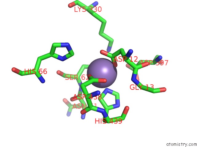
Mono view
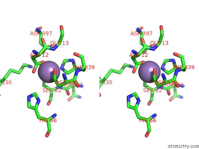
Stereo pair view
Mono view
Stereo pair view
A full contact list of Manganese with other atoms in the Mn binding
site number 1 of Crystal Structure of Phosphoglycerate Mutase From Staphylococcus Aureus in 2-Phosphoglyceric Acid Bound Form within 5.0Å range:
|
Manganese binding site 2 out of 2 in 4nwx
Go back to
Manganese binding site 2 out
of 2 in the Crystal Structure of Phosphoglycerate Mutase From Staphylococcus Aureus in 2-Phosphoglyceric Acid Bound Form
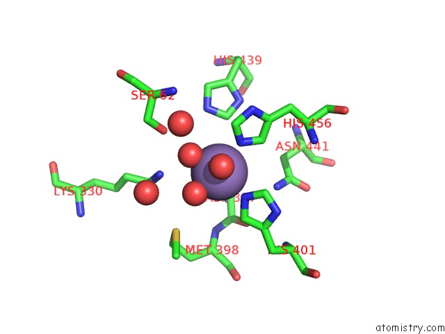
Mono view
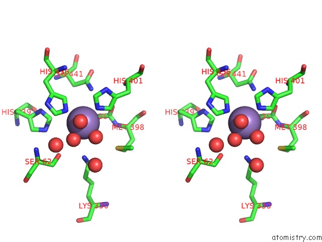
Stereo pair view
Mono view
Stereo pair view
A full contact list of Manganese with other atoms in the Mn binding
site number 2 of Crystal Structure of Phosphoglycerate Mutase From Staphylococcus Aureus in 2-Phosphoglyceric Acid Bound Form within 5.0Å range:
|
Reference:
A.Roychowdhury,
A.Kundu,
M.Bose,
A.Gujar,
S.Mukherjee,
A.K.Das.
Complete Catalytic Cycle of Cofactor-Independent Phosphoglycerate Mutase Involves A Spring-Loaded Mechanism Febs J. 2015.
ISSN: ISSN 1742-464X
PubMed: 25611430
DOI: 10.1111/FEBS.13205
Page generated: Sat Oct 5 20:35:49 2024
ISSN: ISSN 1742-464X
PubMed: 25611430
DOI: 10.1111/FEBS.13205
Last articles
Zn in 9MJ5Zn in 9HNW
Zn in 9G0L
Zn in 9FNE
Zn in 9DZN
Zn in 9E0I
Zn in 9D32
Zn in 9DAK
Zn in 8ZXC
Zn in 8ZUF