Manganese »
PDB 4mu4-4nx7 »
4ni8 »
Manganese in PDB 4ni8: Crystal Structure of 5-Carboxyvanillate Decarboxylase Ligw From Sphingomonas Paucimobilis Complexed with Mn and 5-Methoxyisophtalic Acid
Protein crystallography data
The structure of Crystal Structure of 5-Carboxyvanillate Decarboxylase Ligw From Sphingomonas Paucimobilis Complexed with Mn and 5-Methoxyisophtalic Acid, PDB code: 4ni8
was solved by
A.A.Fedorov,
E.V.Fedorov,
A.Vladimirova,
F.M.Raushel,
S.C.Almo,
with X-Ray Crystallography technique. A brief refinement statistics is given in the table below:
| Resolution Low / High (Å) | 39.97 / 1.64 |
| Space group | P 1 |
| Cell size a, b, c (Å), α, β, γ (°) | 80.523, 99.896, 100.427, 67.04, 88.44, 68.07 |
| R / Rfree (%) | 14.8 / 17.5 |
Manganese Binding Sites:
Pages:
>>> Page 1 <<< Page 2, Binding sites: 11 - 16;Binding sites:
The binding sites of Manganese atom in the Crystal Structure of 5-Carboxyvanillate Decarboxylase Ligw From Sphingomonas Paucimobilis Complexed with Mn and 5-Methoxyisophtalic Acid (pdb code 4ni8). This binding sites where shown within 5.0 Angstroms radius around Manganese atom.In total 16 binding sites of Manganese where determined in the Crystal Structure of 5-Carboxyvanillate Decarboxylase Ligw From Sphingomonas Paucimobilis Complexed with Mn and 5-Methoxyisophtalic Acid, PDB code: 4ni8:
Jump to Manganese binding site number: 1; 2; 3; 4; 5; 6; 7; 8; 9; 10;
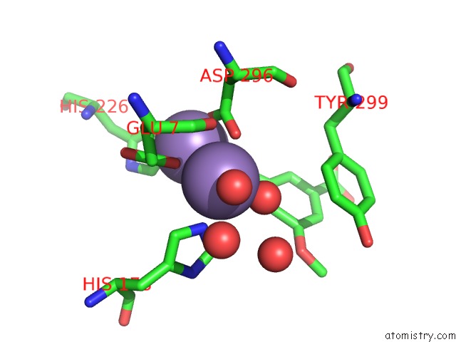
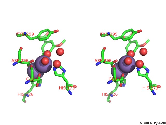
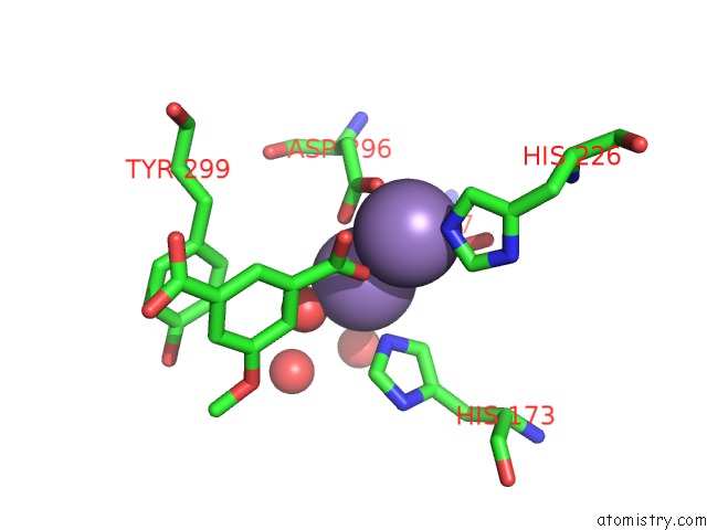
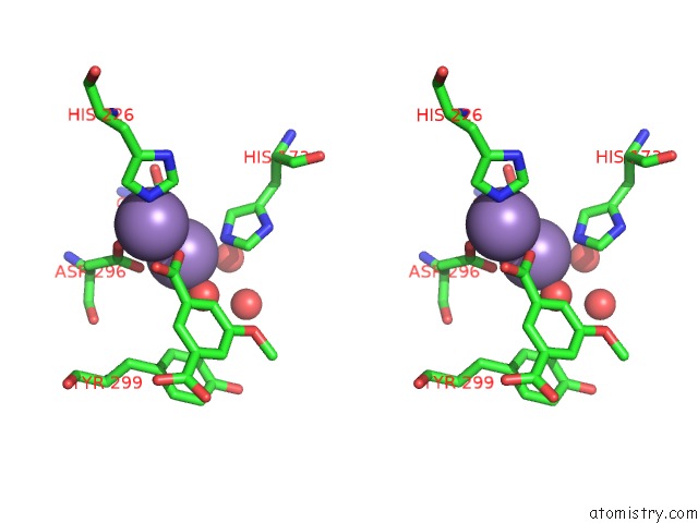
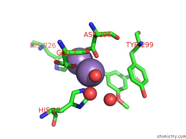
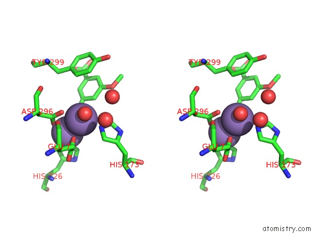
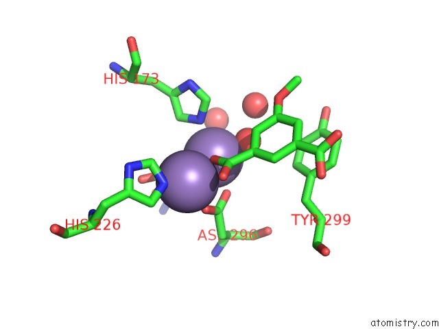
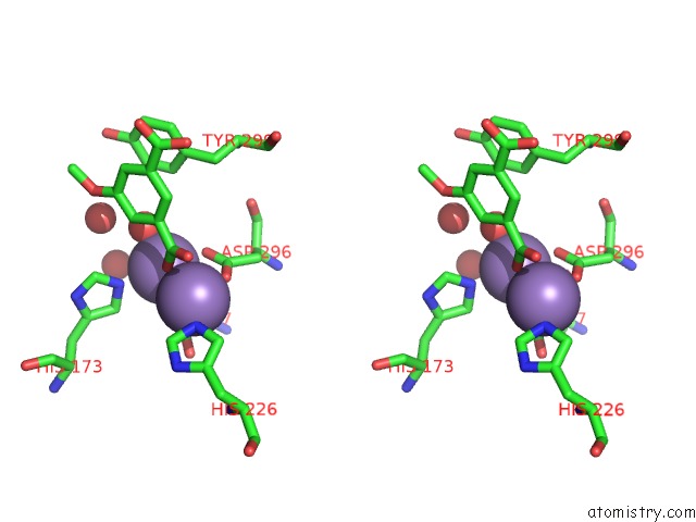
Manganese binding site 1 out of 16 in 4ni8
Go back to
Manganese binding site 1 out
of 16 in the Crystal Structure of 5-Carboxyvanillate Decarboxylase Ligw From Sphingomonas Paucimobilis Complexed with Mn and 5-Methoxyisophtalic Acid
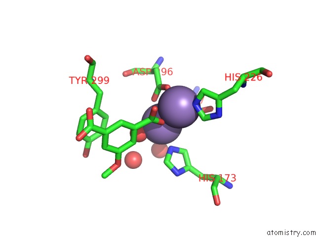
Mono view
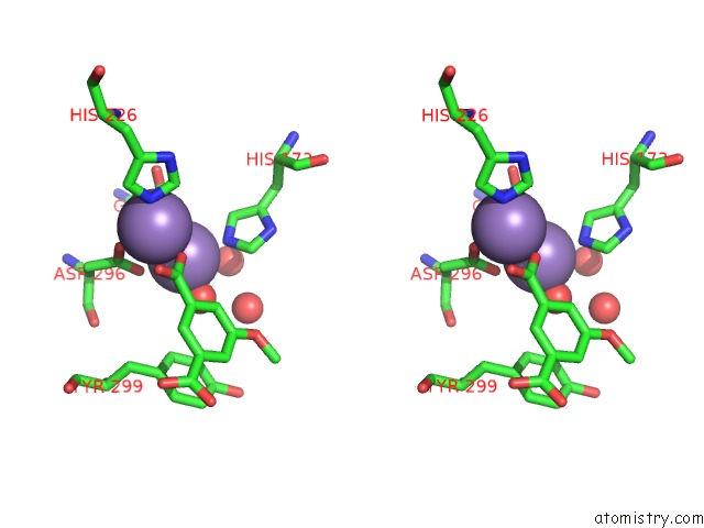
Stereo pair view
Mono view
Stereo pair view
A full contact list of Manganese with other atoms in the Mn binding
site number 1 of Crystal Structure of 5-Carboxyvanillate Decarboxylase Ligw From Sphingomonas Paucimobilis Complexed with Mn and 5-Methoxyisophtalic Acid within 5.0Å range:
|
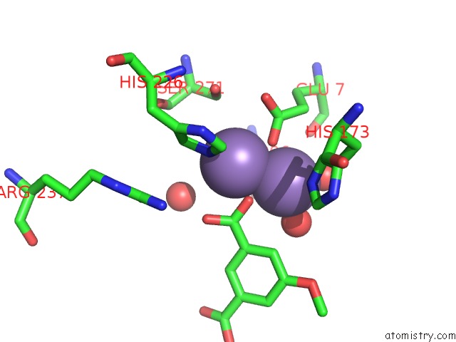
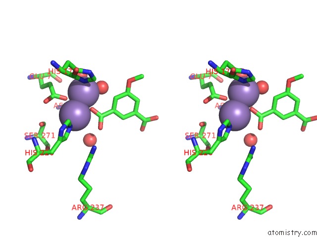
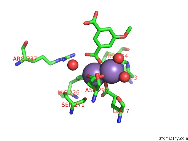
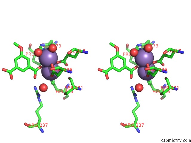
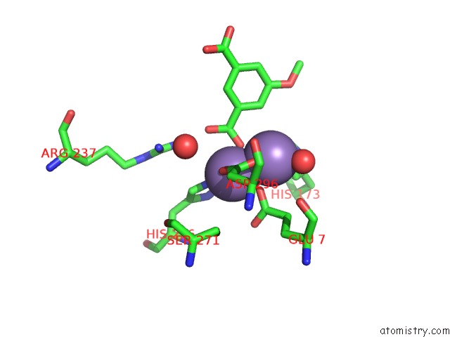
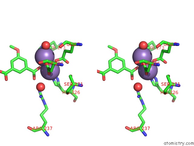
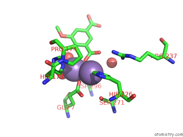
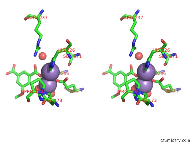
Manganese binding site 2 out of 16 in 4ni8
Go back to
Manganese binding site 2 out
of 16 in the Crystal Structure of 5-Carboxyvanillate Decarboxylase Ligw From Sphingomonas Paucimobilis Complexed with Mn and 5-Methoxyisophtalic Acid
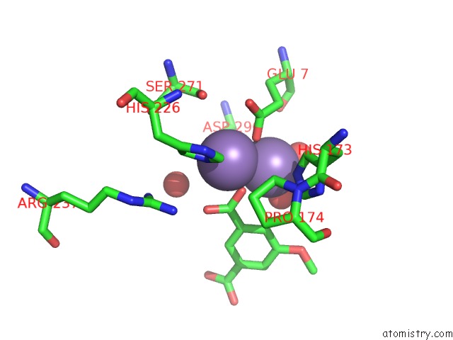
Mono view
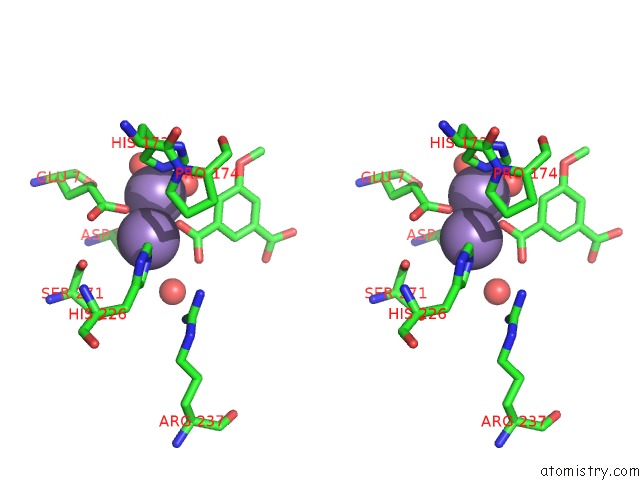
Stereo pair view
Mono view
Stereo pair view
A full contact list of Manganese with other atoms in the Mn binding
site number 2 of Crystal Structure of 5-Carboxyvanillate Decarboxylase Ligw From Sphingomonas Paucimobilis Complexed with Mn and 5-Methoxyisophtalic Acid within 5.0Å range:
|
Manganese binding site 3 out of 16 in 4ni8
Go back to
Manganese binding site 3 out
of 16 in the Crystal Structure of 5-Carboxyvanillate Decarboxylase Ligw From Sphingomonas Paucimobilis Complexed with Mn and 5-Methoxyisophtalic Acid
Mono view
Stereo pair view
Mono view
Stereo pair view
A full contact list of Manganese with other atoms in the Mn binding
site number 3 of Crystal Structure of 5-Carboxyvanillate Decarboxylase Ligw From Sphingomonas Paucimobilis Complexed with Mn and 5-Methoxyisophtalic Acid within 5.0Å range:
|
Manganese binding site 4 out of 16 in 4ni8
Go back to
Manganese binding site 4 out
of 16 in the Crystal Structure of 5-Carboxyvanillate Decarboxylase Ligw From Sphingomonas Paucimobilis Complexed with Mn and 5-Methoxyisophtalic Acid
Mono view
Stereo pair view
Mono view
Stereo pair view
A full contact list of Manganese with other atoms in the Mn binding
site number 4 of Crystal Structure of 5-Carboxyvanillate Decarboxylase Ligw From Sphingomonas Paucimobilis Complexed with Mn and 5-Methoxyisophtalic Acid within 5.0Å range:
|
Manganese binding site 5 out of 16 in 4ni8
Go back to
Manganese binding site 5 out
of 16 in the Crystal Structure of 5-Carboxyvanillate Decarboxylase Ligw From Sphingomonas Paucimobilis Complexed with Mn and 5-Methoxyisophtalic Acid
Mono view
Stereo pair view
Mono view
Stereo pair view
A full contact list of Manganese with other atoms in the Mn binding
site number 5 of Crystal Structure of 5-Carboxyvanillate Decarboxylase Ligw From Sphingomonas Paucimobilis Complexed with Mn and 5-Methoxyisophtalic Acid within 5.0Å range:
|
Manganese binding site 6 out of 16 in 4ni8
Go back to
Manganese binding site 6 out
of 16 in the Crystal Structure of 5-Carboxyvanillate Decarboxylase Ligw From Sphingomonas Paucimobilis Complexed with Mn and 5-Methoxyisophtalic Acid
Mono view
Stereo pair view
Mono view
Stereo pair view
A full contact list of Manganese with other atoms in the Mn binding
site number 6 of Crystal Structure of 5-Carboxyvanillate Decarboxylase Ligw From Sphingomonas Paucimobilis Complexed with Mn and 5-Methoxyisophtalic Acid within 5.0Å range:
|
Manganese binding site 7 out of 16 in 4ni8
Go back to
Manganese binding site 7 out
of 16 in the Crystal Structure of 5-Carboxyvanillate Decarboxylase Ligw From Sphingomonas Paucimobilis Complexed with Mn and 5-Methoxyisophtalic Acid
Mono view
Stereo pair view
Mono view
Stereo pair view
A full contact list of Manganese with other atoms in the Mn binding
site number 7 of Crystal Structure of 5-Carboxyvanillate Decarboxylase Ligw From Sphingomonas Paucimobilis Complexed with Mn and 5-Methoxyisophtalic Acid within 5.0Å range:
|
Manganese binding site 8 out of 16 in 4ni8
Go back to
Manganese binding site 8 out
of 16 in the Crystal Structure of 5-Carboxyvanillate Decarboxylase Ligw From Sphingomonas Paucimobilis Complexed with Mn and 5-Methoxyisophtalic Acid
Mono view
Stereo pair view
Mono view
Stereo pair view
A full contact list of Manganese with other atoms in the Mn binding
site number 8 of Crystal Structure of 5-Carboxyvanillate Decarboxylase Ligw From Sphingomonas Paucimobilis Complexed with Mn and 5-Methoxyisophtalic Acid within 5.0Å range:
|
Manganese binding site 9 out of 16 in 4ni8
Go back to
Manganese binding site 9 out
of 16 in the Crystal Structure of 5-Carboxyvanillate Decarboxylase Ligw From Sphingomonas Paucimobilis Complexed with Mn and 5-Methoxyisophtalic Acid
Mono view
Stereo pair view
Mono view
Stereo pair view
A full contact list of Manganese with other atoms in the Mn binding
site number 9 of Crystal Structure of 5-Carboxyvanillate Decarboxylase Ligw From Sphingomonas Paucimobilis Complexed with Mn and 5-Methoxyisophtalic Acid within 5.0Å range:
|
Manganese binding site 10 out of 16 in 4ni8
Go back to
Manganese binding site 10 out
of 16 in the Crystal Structure of 5-Carboxyvanillate Decarboxylase Ligw From Sphingomonas Paucimobilis Complexed with Mn and 5-Methoxyisophtalic Acid
Mono view
Stereo pair view
Mono view
Stereo pair view
A full contact list of Manganese with other atoms in the Mn binding
site number 10 of Crystal Structure of 5-Carboxyvanillate Decarboxylase Ligw From Sphingomonas Paucimobilis Complexed with Mn and 5-Methoxyisophtalic Acid within 5.0Å range:
|
Reference:
A.A.Fedorov,
E.V.Fedorov,
A.Vladimirova,
F.M.Raushel,
S.C.Almo.
Crystal Structure of 5-Carboxyvanillate Decarboxylase Ligw From Sphingomonas Paucimobilis Complexed with Mn and 5-Methoxyisophtalic Acid To Be Published.
Page generated: Sat Oct 5 20:33:03 2024
Last articles
Cl in 3DHKCl in 3DHH
Cl in 3DCI
Cl in 3DGC
Cl in 3DGO
Cl in 3DFJ
Cl in 3DGA
Cl in 3DEW
Cl in 3DEP
Cl in 3DEJ