Manganese »
PDB 4mu4-4nx7 »
4n83 »
Manganese in PDB 4n83: X-Ray Crystal Structure of Streptococcus Sanguinis Dimanganese(II)- Nrdf
Enzymatic activity of X-Ray Crystal Structure of Streptococcus Sanguinis Dimanganese(II)- Nrdf
All present enzymatic activity of X-Ray Crystal Structure of Streptococcus Sanguinis Dimanganese(II)- Nrdf:
1.17.4.1;
1.17.4.1;
Protein crystallography data
The structure of X-Ray Crystal Structure of Streptococcus Sanguinis Dimanganese(II)- Nrdf, PDB code: 4n83
was solved by
A.K.Boal,
A.C.Rosenzweig,
with X-Ray Crystallography technique. A brief refinement statistics is given in the table below:
| Resolution Low / High (Å) | 29.90 / 2.65 |
| Space group | P 1 21 1 |
| Cell size a, b, c (Å), α, β, γ (°) | 117.657, 80.224, 166.374, 90.00, 105.91, 90.00 |
| R / Rfree (%) | 23.5 / 27.8 |
Manganese Binding Sites:
Pages:
>>> Page 1 <<< Page 2, Binding sites: 11 - 16;Binding sites:
The binding sites of Manganese atom in the X-Ray Crystal Structure of Streptococcus Sanguinis Dimanganese(II)- Nrdf (pdb code 4n83). This binding sites where shown within 5.0 Angstroms radius around Manganese atom.In total 16 binding sites of Manganese where determined in the X-Ray Crystal Structure of Streptococcus Sanguinis Dimanganese(II)- Nrdf, PDB code: 4n83:
Jump to Manganese binding site number: 1; 2; 3; 4; 5; 6; 7; 8; 9; 10;
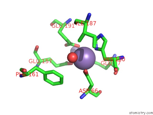
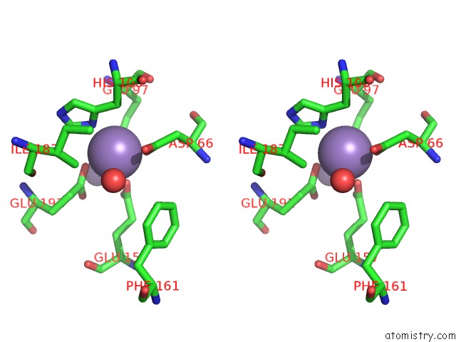
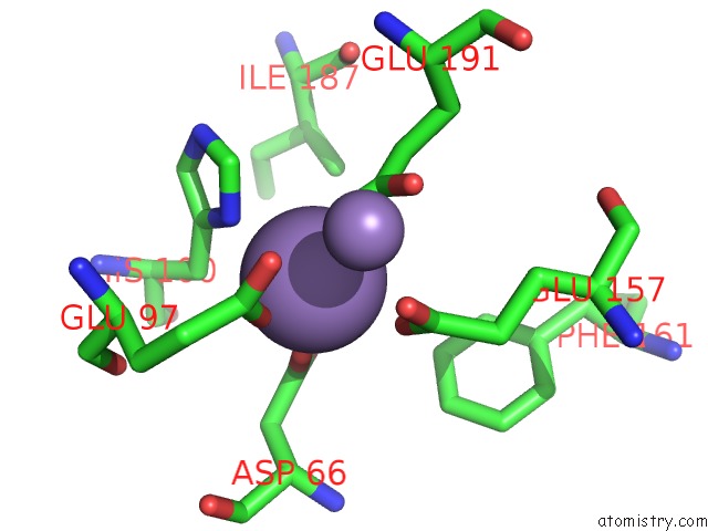
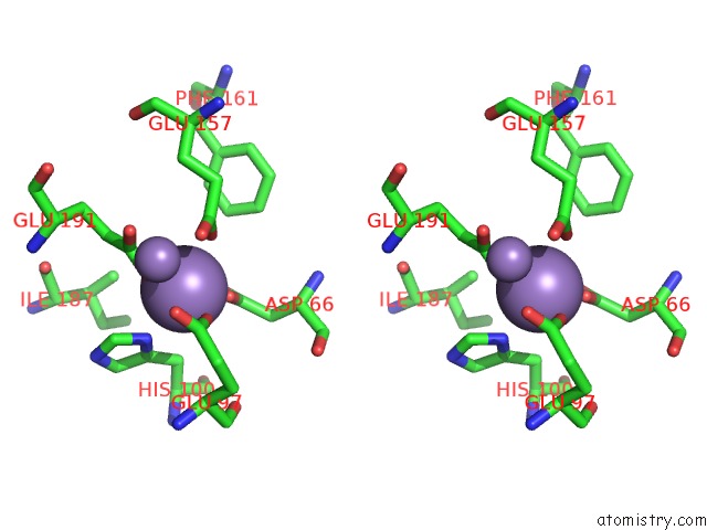
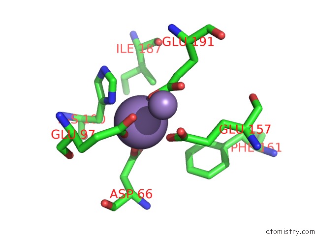
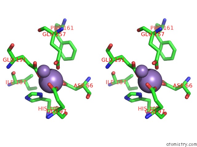
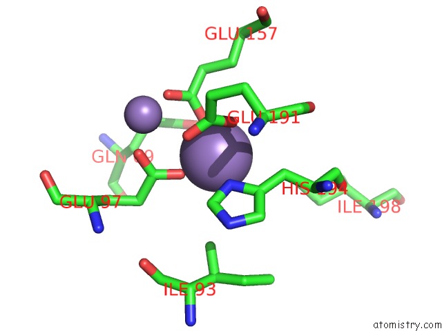
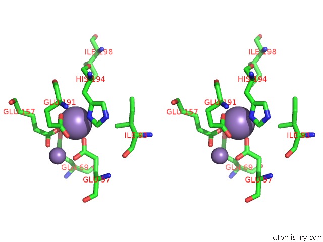
Manganese binding site 1 out of 16 in 4n83
Go back to
Manganese binding site 1 out
of 16 in the X-Ray Crystal Structure of Streptococcus Sanguinis Dimanganese(II)- Nrdf
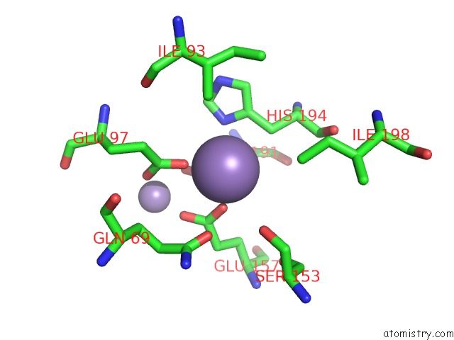
Mono view
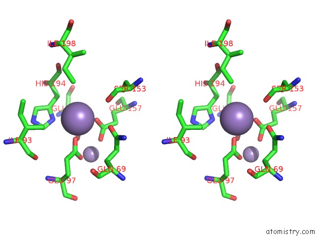
Stereo pair view
Mono view
Stereo pair view
A full contact list of Manganese with other atoms in the Mn binding
site number 1 of X-Ray Crystal Structure of Streptococcus Sanguinis Dimanganese(II)- Nrdf within 5.0Å range:
|
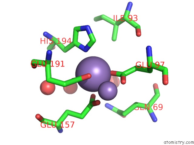
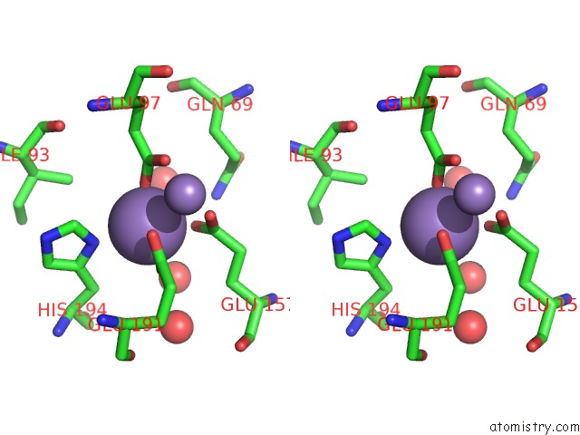
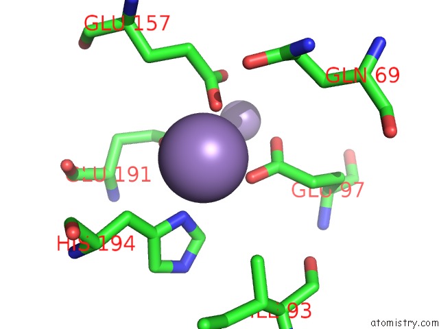
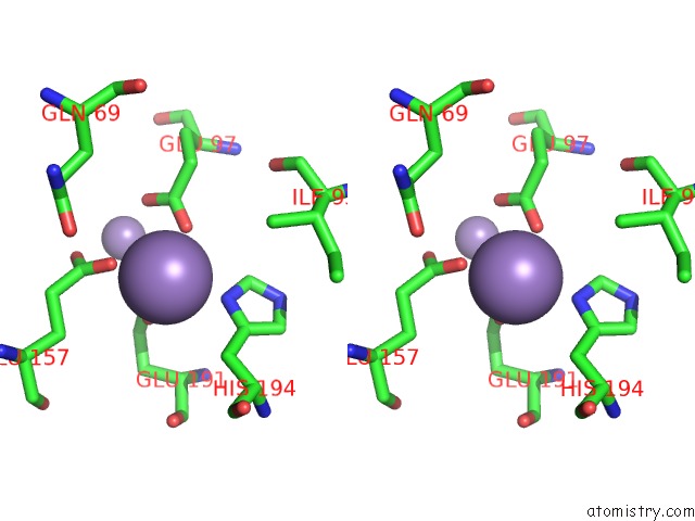
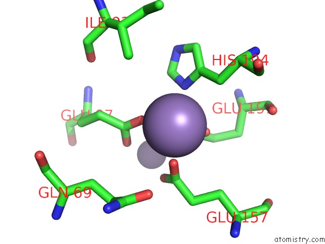
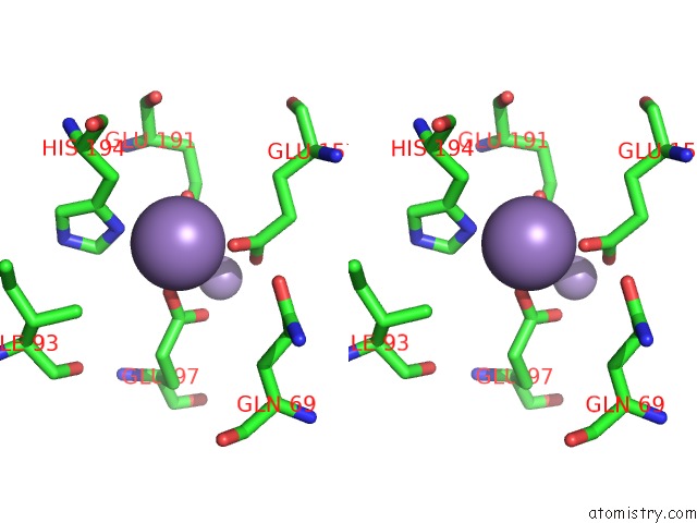
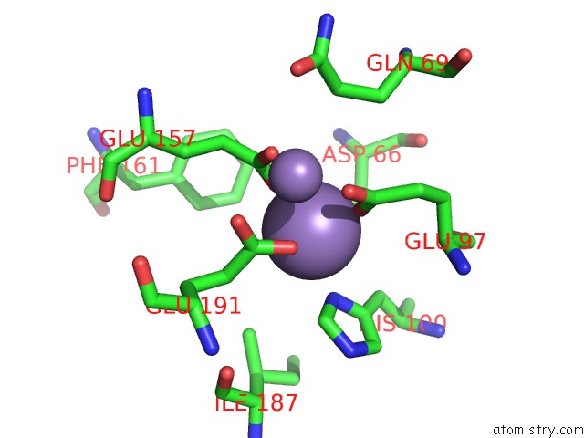
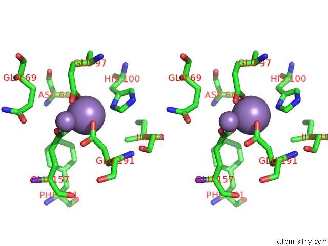
Manganese binding site 2 out of 16 in 4n83
Go back to
Manganese binding site 2 out
of 16 in the X-Ray Crystal Structure of Streptococcus Sanguinis Dimanganese(II)- Nrdf
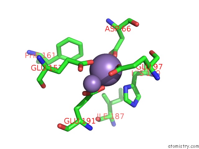
Mono view
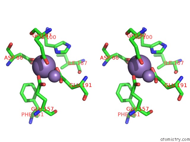
Stereo pair view
Mono view
Stereo pair view
A full contact list of Manganese with other atoms in the Mn binding
site number 2 of X-Ray Crystal Structure of Streptococcus Sanguinis Dimanganese(II)- Nrdf within 5.0Å range:
|
Manganese binding site 3 out of 16 in 4n83
Go back to
Manganese binding site 3 out
of 16 in the X-Ray Crystal Structure of Streptococcus Sanguinis Dimanganese(II)- Nrdf
Mono view
Stereo pair view
Mono view
Stereo pair view
A full contact list of Manganese with other atoms in the Mn binding
site number 3 of X-Ray Crystal Structure of Streptococcus Sanguinis Dimanganese(II)- Nrdf within 5.0Å range:
|
Manganese binding site 4 out of 16 in 4n83
Go back to
Manganese binding site 4 out
of 16 in the X-Ray Crystal Structure of Streptococcus Sanguinis Dimanganese(II)- Nrdf
Mono view
Stereo pair view
Mono view
Stereo pair view
A full contact list of Manganese with other atoms in the Mn binding
site number 4 of X-Ray Crystal Structure of Streptococcus Sanguinis Dimanganese(II)- Nrdf within 5.0Å range:
|
Manganese binding site 5 out of 16 in 4n83
Go back to
Manganese binding site 5 out
of 16 in the X-Ray Crystal Structure of Streptococcus Sanguinis Dimanganese(II)- Nrdf
Mono view
Stereo pair view
Mono view
Stereo pair view
A full contact list of Manganese with other atoms in the Mn binding
site number 5 of X-Ray Crystal Structure of Streptococcus Sanguinis Dimanganese(II)- Nrdf within 5.0Å range:
|
Manganese binding site 6 out of 16 in 4n83
Go back to
Manganese binding site 6 out
of 16 in the X-Ray Crystal Structure of Streptococcus Sanguinis Dimanganese(II)- Nrdf
Mono view
Stereo pair view
Mono view
Stereo pair view
A full contact list of Manganese with other atoms in the Mn binding
site number 6 of X-Ray Crystal Structure of Streptococcus Sanguinis Dimanganese(II)- Nrdf within 5.0Å range:
|
Manganese binding site 7 out of 16 in 4n83
Go back to
Manganese binding site 7 out
of 16 in the X-Ray Crystal Structure of Streptococcus Sanguinis Dimanganese(II)- Nrdf
Mono view
Stereo pair view
Mono view
Stereo pair view
A full contact list of Manganese with other atoms in the Mn binding
site number 7 of X-Ray Crystal Structure of Streptococcus Sanguinis Dimanganese(II)- Nrdf within 5.0Å range:
|
Manganese binding site 8 out of 16 in 4n83
Go back to
Manganese binding site 8 out
of 16 in the X-Ray Crystal Structure of Streptococcus Sanguinis Dimanganese(II)- Nrdf
Mono view
Stereo pair view
Mono view
Stereo pair view
A full contact list of Manganese with other atoms in the Mn binding
site number 8 of X-Ray Crystal Structure of Streptococcus Sanguinis Dimanganese(II)- Nrdf within 5.0Å range:
|
Manganese binding site 9 out of 16 in 4n83
Go back to
Manganese binding site 9 out
of 16 in the X-Ray Crystal Structure of Streptococcus Sanguinis Dimanganese(II)- Nrdf
Mono view
Stereo pair view
Mono view
Stereo pair view
A full contact list of Manganese with other atoms in the Mn binding
site number 9 of X-Ray Crystal Structure of Streptococcus Sanguinis Dimanganese(II)- Nrdf within 5.0Å range:
|
Manganese binding site 10 out of 16 in 4n83
Go back to
Manganese binding site 10 out
of 16 in the X-Ray Crystal Structure of Streptococcus Sanguinis Dimanganese(II)- Nrdf
Mono view
Stereo pair view
Mono view
Stereo pair view
A full contact list of Manganese with other atoms in the Mn binding
site number 10 of X-Ray Crystal Structure of Streptococcus Sanguinis Dimanganese(II)- Nrdf within 5.0Å range:
|
Reference:
O.Makhlynets,
A.K.Boal,
D.V.Rhodes,
T.Kitten,
A.C.Rosenzweig,
J.Stubbe.
Streptococcus Sanguinis Class Ib Ribonucleotide Reductase: High Activity with Both Iron and Manganese Cofactors and Structural Insights. J.Biol.Chem. V. 289 6259 2014.
ISSN: ISSN 0021-9258
PubMed: 24381172
DOI: 10.1074/JBC.M113.533554
Page generated: Sat Oct 5 20:30:13 2024
ISSN: ISSN 0021-9258
PubMed: 24381172
DOI: 10.1074/JBC.M113.533554
Last articles
Zn in 9MJ5Zn in 9HNW
Zn in 9G0L
Zn in 9FNE
Zn in 9DZN
Zn in 9E0I
Zn in 9D32
Zn in 9DAK
Zn in 8ZXC
Zn in 8ZUF