Manganese in PDB 4mr0: Crystal Structure of Pfba, A Surface Adhesin of Streptococcus Pneumoniae
Protein crystallography data
The structure of Crystal Structure of Pfba, A Surface Adhesin of Streptococcus Pneumoniae, PDB code: 4mr0
was solved by
K.Ponnuraj,
D.S.J.Beulin,
with X-Ray Crystallography technique. A brief refinement statistics is given in the table below:
Resolution Low / High (Å)
|
20.00 /
1.95
|
Space group
|
P 1 21 1
|
Cell size a, b, c (Å), α, β, γ (°)
|
60.590,
126.080,
63.480,
90.00,
99.60,
90.00
|
R / Rfree (%)
|
20.4 /
22.2
|
Other elements in 4mr0:
The structure of Crystal Structure of Pfba, A Surface Adhesin of Streptococcus Pneumoniae also contains other interesting chemical elements:
Manganese Binding Sites:
The binding sites of Manganese atom in the Crystal Structure of Pfba, A Surface Adhesin of Streptococcus Pneumoniae
(pdb code 4mr0). This binding sites where shown within
5.0 Angstroms radius around Manganese atom.
In total 8 binding sites of Manganese where determined in the
Crystal Structure of Pfba, A Surface Adhesin of Streptococcus Pneumoniae, PDB code: 4mr0:
Jump to Manganese binding site number:
1;
2;
3;
4;
5;
6;
7;
8;
Manganese binding site 1 out
of 8 in 4mr0
Go back to
Manganese Binding Sites List in 4mr0
Manganese binding site 1 out
of 8 in the Crystal Structure of Pfba, A Surface Adhesin of Streptococcus Pneumoniae
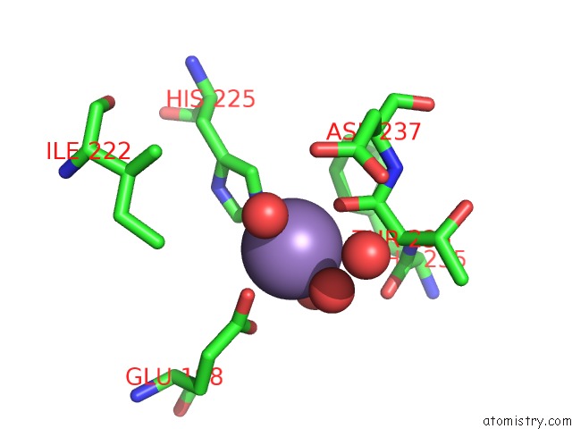
Mono view
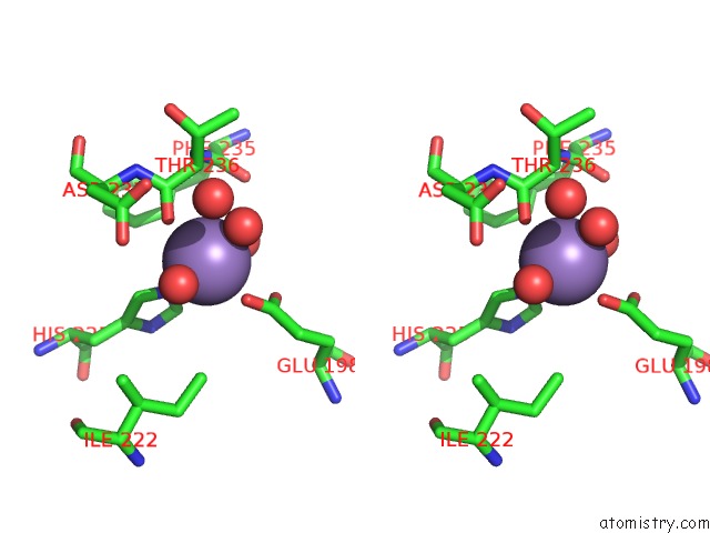
Stereo pair view
|
A full contact list of Manganese with other atoms in the Mn binding
site number 1 of Crystal Structure of Pfba, A Surface Adhesin of Streptococcus Pneumoniae within 5.0Å range:
probe
|
atom
|
residue
|
distance (Å)
|
B
|
Occ
|
A:Mn704
b:19.5
occ:1.00
|
OE2
|
A:GLU198
|
2.2
|
18.6
|
1.0
|
O
|
A:THR236
|
2.2
|
16.0
|
1.0
|
O
|
A:HOH1026
|
2.3
|
21.7
|
1.0
|
NE2
|
A:HIS225
|
2.3
|
15.5
|
1.0
|
O
|
A:HOH974
|
2.4
|
19.2
|
1.0
|
O
|
A:HOH1027
|
2.4
|
14.8
|
1.0
|
CD2
|
A:HIS225
|
3.2
|
14.9
|
1.0
|
CD
|
A:GLU198
|
3.2
|
19.4
|
1.0
|
C
|
A:THR236
|
3.3
|
18.1
|
1.0
|
CE1
|
A:HIS225
|
3.4
|
15.0
|
1.0
|
OE1
|
A:GLU198
|
3.5
|
19.1
|
1.0
|
CA
|
A:THR236
|
3.8
|
16.7
|
1.0
|
OD2
|
A:ASP237
|
4.3
|
23.1
|
1.0
|
O
|
A:HOH846
|
4.3
|
22.9
|
1.0
|
O
|
A:PHE235
|
4.4
|
18.0
|
1.0
|
CG
|
A:HIS225
|
4.4
|
16.4
|
1.0
|
ND1
|
A:HIS225
|
4.4
|
15.7
|
1.0
|
CD1
|
A:PHE235
|
4.4
|
17.3
|
1.0
|
N
|
A:ASP237
|
4.5
|
18.7
|
1.0
|
CD1
|
A:ILE222
|
4.6
|
19.9
|
1.0
|
CG
|
A:GLU198
|
4.6
|
16.5
|
1.0
|
N
|
A:THR236
|
4.6
|
17.4
|
1.0
|
CG
|
A:ASP237
|
4.6
|
22.2
|
1.0
|
CE1
|
A:PHE235
|
4.6
|
17.6
|
1.0
|
OD1
|
A:ASP237
|
4.8
|
23.0
|
1.0
|
C
|
A:PHE235
|
4.8
|
17.8
|
1.0
|
CA
|
A:ASP237
|
4.9
|
19.1
|
1.0
|
CB
|
A:THR236
|
5.0
|
18.2
|
1.0
|
|
Manganese binding site 2 out
of 8 in 4mr0
Go back to
Manganese Binding Sites List in 4mr0
Manganese binding site 2 out
of 8 in the Crystal Structure of Pfba, A Surface Adhesin of Streptococcus Pneumoniae
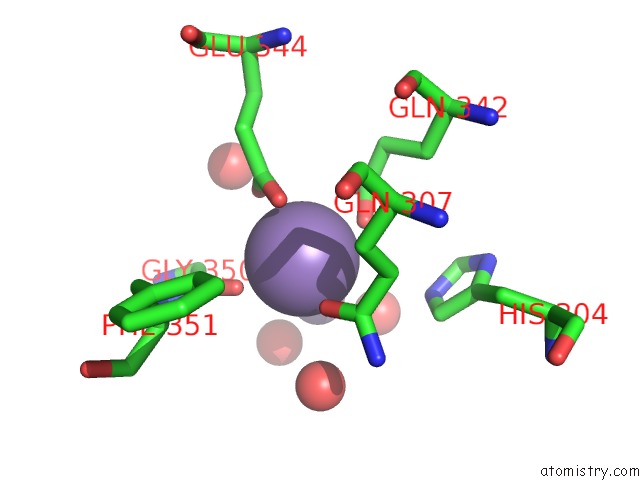
Mono view
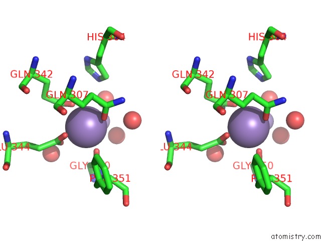
Stereo pair view
|
A full contact list of Manganese with other atoms in the Mn binding
site number 2 of Crystal Structure of Pfba, A Surface Adhesin of Streptococcus Pneumoniae within 5.0Å range:
probe
|
atom
|
residue
|
distance (Å)
|
B
|
Occ
|
A:Mn705
b:23.9
occ:1.00
|
O
|
A:GLY350
|
2.2
|
17.0
|
1.0
|
OE1
|
A:GLN342
|
2.2
|
15.1
|
1.0
|
OE2
|
A:GLU344
|
2.3
|
19.9
|
1.0
|
OE1
|
A:GLN307
|
2.3
|
16.5
|
1.0
|
OE1
|
A:GLU344
|
2.6
|
19.1
|
1.0
|
CD
|
A:GLU344
|
2.8
|
18.2
|
1.0
|
MN
|
A:MN707
|
3.1
|
50.1
|
1.0
|
CD
|
A:GLN342
|
3.1
|
18.8
|
1.0
|
C
|
A:GLY350
|
3.3
|
18.0
|
1.0
|
CD
|
A:GLN307
|
3.4
|
18.6
|
1.0
|
O
|
A:HOH1042
|
3.4
|
54.8
|
1.0
|
NE2
|
A:GLN342
|
3.6
|
18.1
|
1.0
|
CG
|
A:GLN307
|
3.9
|
16.8
|
1.0
|
O
|
A:HOH878
|
3.9
|
30.3
|
1.0
|
CA
|
A:GLY350
|
3.9
|
18.3
|
1.0
|
CG
|
A:GLU344
|
4.3
|
17.8
|
1.0
|
CG
|
A:GLN342
|
4.3
|
15.9
|
1.0
|
N
|
A:PHE351
|
4.4
|
15.4
|
1.0
|
NE2
|
A:HIS304
|
4.4
|
18.8
|
1.0
|
CB
|
A:GLN307
|
4.4
|
15.6
|
1.0
|
CD2
|
A:HIS304
|
4.5
|
18.5
|
1.0
|
NE2
|
A:GLN307
|
4.5
|
16.5
|
1.0
|
O
|
A:HOH1040
|
4.5
|
37.7
|
1.0
|
O
|
A:HOH903
|
4.7
|
14.1
|
1.0
|
CA
|
A:PHE351
|
4.7
|
15.0
|
1.0
|
O
|
A:HOH1076
|
4.8
|
47.8
|
1.0
|
CD1
|
A:PHE351
|
4.8
|
14.9
|
1.0
|
CB
|
A:GLN342
|
4.9
|
14.8
|
1.0
|
|
Manganese binding site 3 out
of 8 in 4mr0
Go back to
Manganese Binding Sites List in 4mr0
Manganese binding site 3 out
of 8 in the Crystal Structure of Pfba, A Surface Adhesin of Streptococcus Pneumoniae
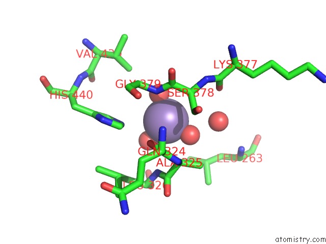
Mono view
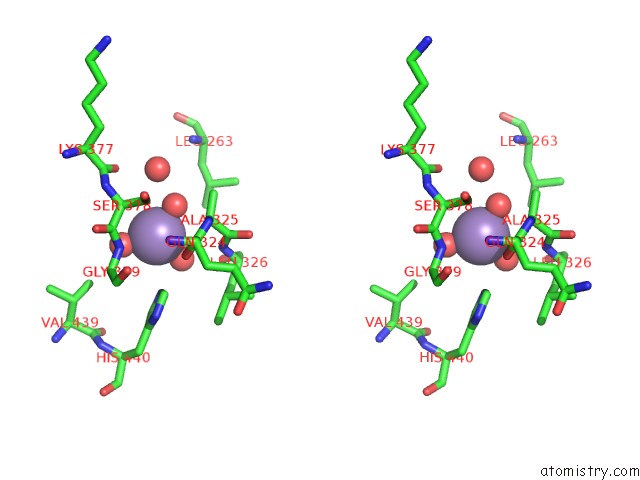
Stereo pair view
|
A full contact list of Manganese with other atoms in the Mn binding
site number 3 of Crystal Structure of Pfba, A Surface Adhesin of Streptococcus Pneumoniae within 5.0Å range:
probe
|
atom
|
residue
|
distance (Å)
|
B
|
Occ
|
A:Mn706
b:46.3
occ:1.00
|
O
|
A:HOH1028
|
2.0
|
31.2
|
1.0
|
O
|
A:HOH1030
|
2.1
|
44.4
|
1.0
|
O
|
A:HOH1029
|
2.1
|
37.8
|
1.0
|
O
|
A:HOH1041
|
2.4
|
42.2
|
1.0
|
NE2
|
A:HIS440
|
3.5
|
26.9
|
1.0
|
N
|
A:GLY379
|
3.7
|
22.6
|
1.0
|
O
|
A:HOH978
|
3.9
|
26.1
|
1.0
|
N
|
A:LEU326
|
4.0
|
26.0
|
1.0
|
CA
|
A:SER378
|
4.1
|
25.2
|
1.0
|
CA
|
A:ALA325
|
4.1
|
22.8
|
1.0
|
CE1
|
A:HIS440
|
4.2
|
25.9
|
1.0
|
CB
|
A:SER378
|
4.3
|
25.6
|
1.0
|
CD1
|
A:LEU263
|
4.3
|
26.5
|
1.0
|
O
|
A:GLN324
|
4.4
|
20.2
|
1.0
|
C
|
A:SER378
|
4.4
|
23.4
|
1.0
|
CD2
|
A:HIS440
|
4.6
|
26.0
|
1.0
|
O
|
A:LEU326
|
4.6
|
31.7
|
1.0
|
C
|
A:ALA325
|
4.6
|
24.5
|
1.0
|
CA
|
A:GLY379
|
4.7
|
21.8
|
1.0
|
CB
|
A:ALA325
|
4.7
|
22.1
|
1.0
|
CG
|
A:LEU263
|
4.7
|
26.3
|
1.0
|
O
|
A:LYS377
|
4.8
|
28.2
|
1.0
|
CG1
|
A:VAL439
|
4.9
|
32.6
|
1.0
|
|
Manganese binding site 4 out
of 8 in 4mr0
Go back to
Manganese Binding Sites List in 4mr0
Manganese binding site 4 out
of 8 in the Crystal Structure of Pfba, A Surface Adhesin of Streptococcus Pneumoniae
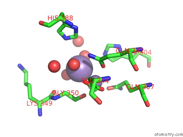
Mono view
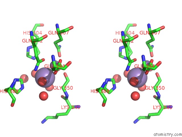
Stereo pair view
|
A full contact list of Manganese with other atoms in the Mn binding
site number 4 of Crystal Structure of Pfba, A Surface Adhesin of Streptococcus Pneumoniae within 5.0Å range:
probe
|
atom
|
residue
|
distance (Å)
|
B
|
Occ
|
A:Mn707
b:50.1
occ:1.00
|
O
|
A:HOH1040
|
2.1
|
37.7
|
1.0
|
O
|
A:HOH1076
|
2.2
|
47.8
|
1.0
|
O
|
A:HOH1042
|
2.2
|
54.8
|
1.0
|
MN
|
A:MN705
|
3.1
|
23.9
|
1.0
|
O
|
A:HOH878
|
3.5
|
30.3
|
1.0
|
O
|
A:GLY350
|
3.7
|
17.0
|
1.0
|
OE1
|
A:GLN342
|
3.7
|
15.1
|
1.0
|
NE2
|
A:GLN342
|
3.8
|
18.1
|
1.0
|
NE2
|
A:HIS304
|
3.8
|
18.8
|
1.0
|
O
|
A:HOH998
|
3.9
|
31.9
|
1.0
|
CA
|
A:GLY350
|
3.9
|
18.3
|
1.0
|
O
|
A:HOH975
|
4.1
|
32.8
|
1.0
|
C
|
A:GLY350
|
4.2
|
18.0
|
1.0
|
CD
|
A:GLN342
|
4.2
|
18.8
|
1.0
|
OE1
|
A:GLU344
|
4.3
|
19.1
|
1.0
|
NE2
|
A:HIS388
|
4.3
|
22.4
|
1.0
|
CE1
|
A:HIS388
|
4.4
|
20.8
|
1.0
|
O
|
A:LYS349
|
4.4
|
19.6
|
1.0
|
OE1
|
A:GLN307
|
4.5
|
16.5
|
1.0
|
CD2
|
A:HIS304
|
4.6
|
18.5
|
1.0
|
CE1
|
A:HIS304
|
4.7
|
19.4
|
1.0
|
|
Manganese binding site 5 out
of 8 in 4mr0
Go back to
Manganese Binding Sites List in 4mr0
Manganese binding site 5 out
of 8 in the Crystal Structure of Pfba, A Surface Adhesin of Streptococcus Pneumoniae
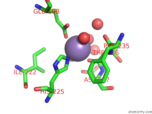
Mono view
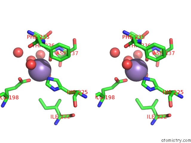
Stereo pair view
|
A full contact list of Manganese with other atoms in the Mn binding
site number 5 of Crystal Structure of Pfba, A Surface Adhesin of Streptococcus Pneumoniae within 5.0Å range:
probe
|
atom
|
residue
|
distance (Å)
|
B
|
Occ
|
B:Mn704
b:18.4
occ:1.00
|
O
|
B:THR236
|
2.2
|
16.4
|
1.0
|
OE1
|
B:GLU198
|
2.2
|
20.2
|
1.0
|
O
|
B:HOH1001
|
2.3
|
19.6
|
1.0
|
O
|
B:HOH1007
|
2.3
|
17.4
|
1.0
|
O
|
B:HOH1009
|
2.3
|
23.5
|
1.0
|
NE2
|
B:HIS225
|
2.4
|
15.6
|
1.0
|
CD2
|
B:HIS225
|
3.2
|
17.4
|
1.0
|
CD
|
B:GLU198
|
3.2
|
21.6
|
1.0
|
C
|
B:THR236
|
3.2
|
17.7
|
1.0
|
CE1
|
B:HIS225
|
3.4
|
16.0
|
1.0
|
OE2
|
B:GLU198
|
3.5
|
19.1
|
1.0
|
CA
|
B:THR236
|
3.7
|
18.7
|
1.0
|
O
|
B:HOH970
|
4.1
|
34.7
|
1.0
|
OD1
|
B:ASP237
|
4.3
|
22.1
|
1.0
|
O
|
B:HOH963
|
4.3
|
26.0
|
1.0
|
O
|
B:PHE235
|
4.3
|
19.2
|
1.0
|
CG
|
B:HIS225
|
4.4
|
17.7
|
1.0
|
N
|
B:ASP237
|
4.4
|
18.5
|
1.0
|
CD2
|
B:PHE235
|
4.4
|
18.6
|
1.0
|
ND1
|
B:HIS225
|
4.5
|
17.4
|
1.0
|
CD1
|
B:ILE222
|
4.5
|
20.6
|
1.0
|
CG
|
B:GLU198
|
4.5
|
18.6
|
1.0
|
N
|
B:THR236
|
4.5
|
17.9
|
1.0
|
CE2
|
B:PHE235
|
4.6
|
20.1
|
1.0
|
CG
|
B:ASP237
|
4.7
|
20.0
|
1.0
|
C
|
B:PHE235
|
4.8
|
18.7
|
1.0
|
CA
|
B:ASP237
|
4.8
|
20.4
|
1.0
|
CB
|
B:THR236
|
4.9
|
19.3
|
1.0
|
OD2
|
B:ASP237
|
4.9
|
20.0
|
1.0
|
|
Manganese binding site 6 out
of 8 in 4mr0
Go back to
Manganese Binding Sites List in 4mr0
Manganese binding site 6 out
of 8 in the Crystal Structure of Pfba, A Surface Adhesin of Streptococcus Pneumoniae
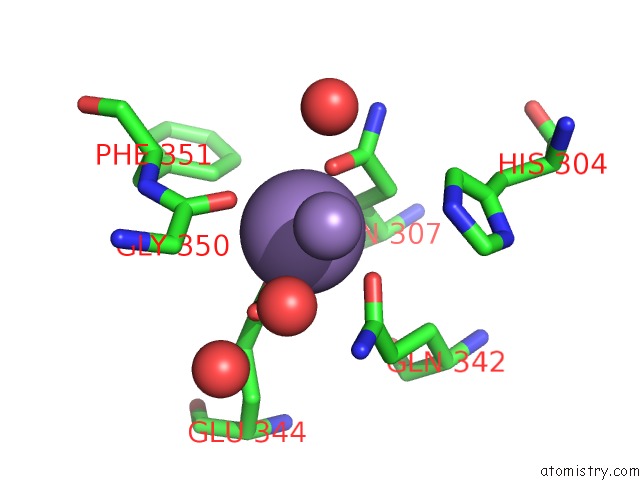
Mono view
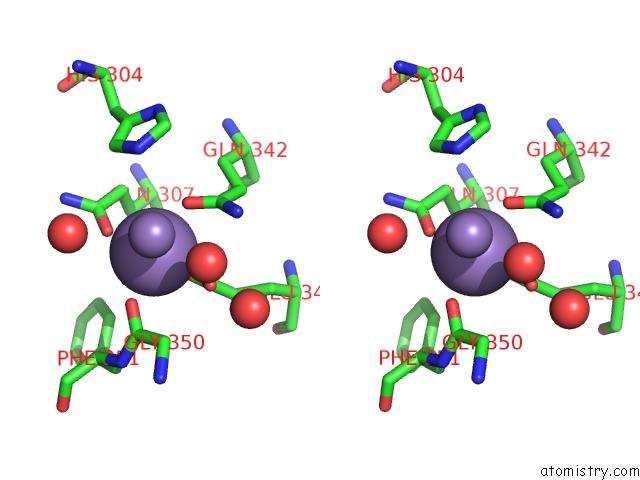
Stereo pair view
|
A full contact list of Manganese with other atoms in the Mn binding
site number 6 of Crystal Structure of Pfba, A Surface Adhesin of Streptococcus Pneumoniae within 5.0Å range:
probe
|
atom
|
residue
|
distance (Å)
|
B
|
Occ
|
B:Mn705
b:28.1
occ:1.00
|
O
|
B:GLY350
|
2.3
|
20.6
|
1.0
|
OE1
|
B:GLN307
|
2.3
|
16.9
|
1.0
|
OE1
|
B:GLN342
|
2.3
|
15.6
|
1.0
|
OE2
|
B:GLU344
|
2.4
|
22.9
|
1.0
|
OE1
|
B:GLU344
|
2.6
|
21.4
|
1.0
|
CD
|
B:GLU344
|
2.8
|
21.4
|
1.0
|
CD
|
B:GLN342
|
3.2
|
18.2
|
1.0
|
MN
|
B:MN707
|
3.3
|
50.1
|
1.0
|
CD
|
B:GLN307
|
3.3
|
19.0
|
1.0
|
C
|
B:GLY350
|
3.4
|
22.6
|
1.0
|
O
|
B:HOH1065
|
3.7
|
33.4
|
1.0
|
NE2
|
B:GLN342
|
3.8
|
21.0
|
1.0
|
O
|
B:HOH1031
|
3.8
|
46.6
|
1.0
|
CG
|
B:GLN307
|
3.9
|
14.5
|
1.0
|
CA
|
B:GLY350
|
4.0
|
24.1
|
1.0
|
CG
|
B:GLU344
|
4.3
|
20.3
|
1.0
|
CG
|
B:GLN342
|
4.3
|
17.9
|
1.0
|
CB
|
B:GLN307
|
4.4
|
14.8
|
1.0
|
NE2
|
B:HIS304
|
4.4
|
17.4
|
1.0
|
CD2
|
B:HIS304
|
4.4
|
20.0
|
1.0
|
N
|
B:PHE351
|
4.4
|
20.4
|
1.0
|
NE2
|
B:GLN307
|
4.5
|
15.7
|
1.0
|
O
|
B:HOH816
|
4.5
|
19.6
|
1.0
|
CA
|
B:PHE351
|
4.7
|
19.3
|
1.0
|
CD1
|
B:PHE351
|
4.8
|
14.2
|
1.0
|
CB
|
B:GLN342
|
4.9
|
15.7
|
1.0
|
|
Manganese binding site 7 out
of 8 in 4mr0
Go back to
Manganese Binding Sites List in 4mr0
Manganese binding site 7 out
of 8 in the Crystal Structure of Pfba, A Surface Adhesin of Streptococcus Pneumoniae
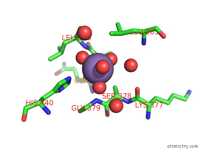
Mono view
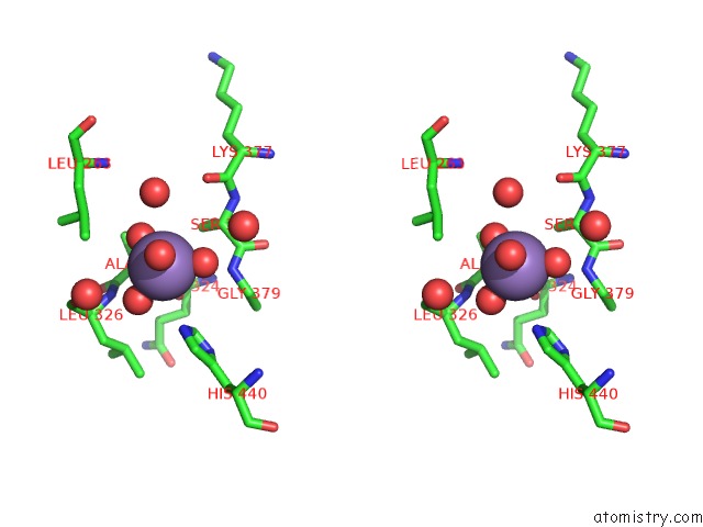
Stereo pair view
|
A full contact list of Manganese with other atoms in the Mn binding
site number 7 of Crystal Structure of Pfba, A Surface Adhesin of Streptococcus Pneumoniae within 5.0Å range:
probe
|
atom
|
residue
|
distance (Å)
|
B
|
Occ
|
B:Mn706
b:39.8
occ:1.00
|
O
|
B:HOH1010
|
2.0
|
29.0
|
1.0
|
O
|
B:HOH1012
|
2.0
|
42.5
|
1.0
|
O
|
B:HOH1011
|
2.1
|
30.5
|
1.0
|
O
|
B:HOH1013
|
2.4
|
30.7
|
1.0
|
NE2
|
B:HIS440
|
3.4
|
21.8
|
1.0
|
O
|
B:HOH962
|
3.8
|
23.6
|
1.0
|
N
|
B:GLY379
|
3.9
|
20.9
|
1.0
|
N
|
B:LEU326
|
3.9
|
25.1
|
1.0
|
CA
|
B:ALA325
|
4.0
|
22.7
|
1.0
|
CE1
|
B:HIS440
|
4.1
|
21.0
|
1.0
|
CD1
|
B:LEU263
|
4.1
|
26.1
|
1.0
|
CA
|
B:SER378
|
4.3
|
22.7
|
1.0
|
O
|
B:GLN324
|
4.3
|
20.0
|
1.0
|
CB
|
B:SER378
|
4.4
|
23.7
|
1.0
|
CD2
|
B:HIS440
|
4.4
|
22.0
|
1.0
|
O
|
B:LEU326
|
4.4
|
28.6
|
1.0
|
C
|
B:ALA325
|
4.5
|
24.2
|
1.0
|
CB
|
B:ALA325
|
4.6
|
22.6
|
1.0
|
C
|
B:SER378
|
4.6
|
22.0
|
1.0
|
O
|
B:HOH924
|
4.6
|
24.8
|
1.0
|
CG
|
B:LEU263
|
4.7
|
26.0
|
1.0
|
O
|
B:HOH859
|
4.7
|
46.2
|
1.0
|
CA
|
B:GLY379
|
4.8
|
21.6
|
1.0
|
CA
|
B:LEU326
|
4.9
|
26.8
|
1.0
|
O
|
B:LYS377
|
5.0
|
26.0
|
1.0
|
|
Manganese binding site 8 out
of 8 in 4mr0
Go back to
Manganese Binding Sites List in 4mr0
Manganese binding site 8 out
of 8 in the Crystal Structure of Pfba, A Surface Adhesin of Streptococcus Pneumoniae
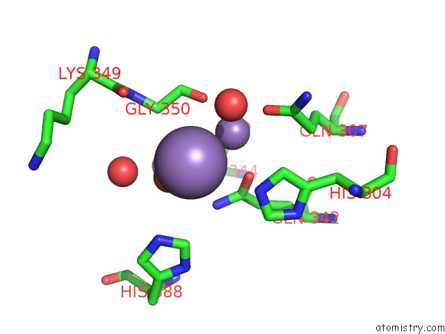
Mono view
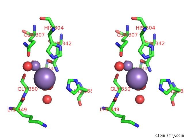
Stereo pair view
|
A full contact list of Manganese with other atoms in the Mn binding
site number 8 of Crystal Structure of Pfba, A Surface Adhesin of Streptococcus Pneumoniae within 5.0Å range:
probe
|
atom
|
residue
|
distance (Å)
|
B
|
Occ
|
B:Mn707
b:50.1
occ:1.00
|
O
|
B:HOH1031
|
2.3
|
46.6
|
1.0
|
MN
|
B:MN705
|
3.3
|
28.1
|
1.0
|
O
|
B:HOH1065
|
3.5
|
33.4
|
1.0
|
NE2
|
B:HIS304
|
3.6
|
17.4
|
1.0
|
OE1
|
B:GLN342
|
3.7
|
15.6
|
1.0
|
NE2
|
B:GLN342
|
3.8
|
21.0
|
1.0
|
O
|
B:GLY350
|
3.9
|
20.6
|
1.0
|
NE2
|
B:HIS388
|
4.0
|
25.1
|
1.0
|
CE1
|
B:HIS388
|
4.1
|
24.3
|
1.0
|
O
|
B:HOH1032
|
4.2
|
39.5
|
1.0
|
CD
|
B:GLN342
|
4.2
|
18.2
|
1.0
|
CA
|
B:GLY350
|
4.2
|
24.1
|
1.0
|
CD2
|
B:HIS304
|
4.4
|
20.0
|
1.0
|
CE1
|
B:HIS304
|
4.5
|
20.6
|
1.0
|
C
|
B:GLY350
|
4.5
|
22.6
|
1.0
|
OE1
|
B:GLU344
|
4.5
|
21.4
|
1.0
|
OE1
|
B:GLN307
|
4.6
|
16.9
|
1.0
|
O
|
B:LYS349
|
4.7
|
29.2
|
1.0
|
O
|
B:HOH816
|
4.9
|
19.6
|
1.0
|
|
Reference:
D.S.J.Beulin,
M.Yamaguchi,
S.Kawabata,
K.Ponnuraj.
Crystal Structure of Pfba, A Surface Adhesin of Streptococcus Pneumoniae, Provides Hints Into Its Interaction with Fibronectin Int.J.Biol.Macromol. V. 64C 168 2013.
ISSN: ISSN 0141-8130
PubMed: 24321492
DOI: 10.1016/J.IJBIOMAC.2013.11.035
Page generated: Sat Oct 5 20:25:11 2024
|