Atomistry » Manganese » PDB 4lta-4mu3 » 4mov
Atomistry »
  Manganese »
    PDB 4lta-4mu3 »
      4mov »

Manganese in PDB 4mov: 1.45 A Resolution Crystal Structure of Protein Phosphatase 1

Enzymatic activity of 1.45 A Resolution Crystal Structure of Protein Phosphatase 1

All present enzymatic activity of 1.45 A Resolution Crystal Structure of Protein Phosphatase 1:
3.1.3.16;

Protein crystallography data

The structure of 1.45 A Resolution Crystal Structure of Protein Phosphatase 1, PDB code: 4mov was solved by M.S.Choy, W.Peti, R.Page, with X-Ray Crystallography technique. A brief refinement statistics is given in the table below:

Resolution Low / High (Å) 46.93 / 1.45
Space group P 21 21 21
Cell size a, b, c (Å), α, β, γ (°) 65.724, 77.600, 133.035, 90.00, 90.00, 90.00
R / Rfree (%) 15 / 16.7

Other elements in 4mov:

The structure of 1.45 A Resolution Crystal Structure of Protein Phosphatase 1 also contains other interesting chemical elements:

Chlorine (Cl) 2 atoms

Manganese Binding Sites:

The binding sites of Manganese atom in the 1.45 A Resolution Crystal Structure of Protein Phosphatase 1 (pdb code 4mov). This binding sites where shown within 5.0 Angstroms radius around Manganese atom.
In total 4 binding sites of Manganese where determined in the 1.45 A Resolution Crystal Structure of Protein Phosphatase 1, PDB code: 4mov:
Jump to Manganese binding site number: 1; 2; 3; 4;

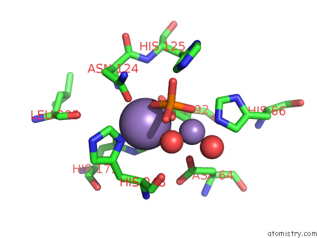
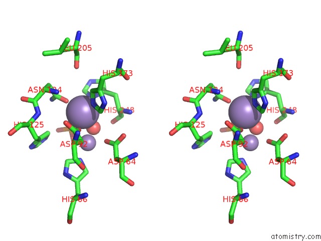
Manganese binding site 1 out of 4 in 4mov

Go back to Manganese Binding Sites List in 4mov
Manganese binding site 1 out of 4 in the 1.45 A Resolution Crystal Structure of Protein Phosphatase 1


Mono view


Stereo pair view

A full contact list of Manganese with other atoms in the Mn binding site number 1 of 1.45 A Resolution Crystal Structure of Protein Phosphatase 1 within 5.0Å range:
probe atom residue distance (Å) B Occ
A:Mn401

b:9.0
occ:1.00
OD1 A:ASN124 2.1 7.9 1.0
O3 A:PO4403 2.2 11.4 0.8
NE2 A:HIS173 2.2 7.7 1.0
OD2 A:ASP92 2.2 8.2 1.0
ND1 A:HIS248 2.2 10.6 1.0
O A:HOH737 2.3 13.0 1.0
CE1 A:HIS248 3.1 11.6 1.0
MN A:MN402 3.1 13.4 0.5
CG A:ASP92 3.2 7.8 1.0
CE1 A:HIS173 3.2 7.3 1.0
CG A:ASN124 3.2 9.2 1.0
CD2 A:HIS173 3.2 6.5 1.0
CG A:HIS248 3.4 9.0 1.0
P A:PO4403 3.4 17.4 0.8
OD1 A:ASP92 3.5 7.5 1.0
ND2 A:ASN124 3.6 10.9 1.0
O1 A:PO4403 3.7 19.6 0.8
CA A:HIS248 3.8 9.9 1.0
CB A:HIS248 3.9 9.5 1.0
OD2 A:ASP64 4.0 11.8 1.0
O4 A:PO4403 4.1 19.7 0.8
O A:HIS248 4.1 17.6 1.0
CD2 A:HIS125 4.2 9.3 1.0
NE2 A:HIS248 4.3 13.5 1.0
ND1 A:HIS173 4.3 6.9 1.0
CG A:HIS173 4.3 7.1 1.0
CD2 A:HIS248 4.4 14.3 1.0
CB A:ASP92 4.4 6.9 1.0
CB A:ASN124 4.5 8.6 1.0
C A:HIS248 4.5 12.0 1.0
N A:ASN124 4.5 7.5 1.0
O2 A:PO4403 4.6 18.8 0.8
O A:LEU205 4.7 10.1 1.0
NE2 A:HIS125 4.8 9.8 1.0
N A:HIS248 4.9 9.5 1.0
NE2 A:HIS66 4.9 9.6 1.0
O A:HOH746 4.9 33.5 1.0
CG A:ASP64 4.9 9.8 1.0

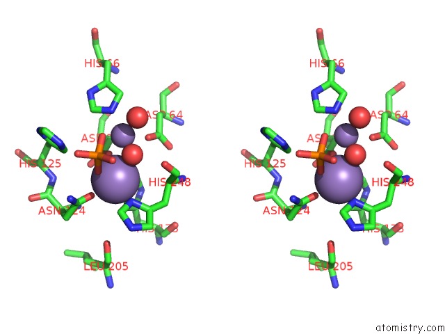
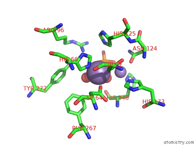
Manganese binding site 2 out of 4 in 4mov

Go back to Manganese Binding Sites List in 4mov
Manganese binding site 2 out of 4 in the 1.45 A Resolution Crystal Structure of Protein Phosphatase 1


Mono view


Stereo pair view

A full contact list of Manganese with other atoms in the Mn binding site number 2 of 1.45 A Resolution Crystal Structure of Protein Phosphatase 1 within 5.0Å range:
probe atom residue distance (Å) B Occ
A:Mn402

b:13.4
occ:0.50
O A:HOH737 1.7 13.0 1.0
NE2 A:HIS66 2.1 9.6 1.0
OD2 A:ASP92 2.2 8.2 1.0
O1 A:PO4403 2.2 19.6 0.8
O A:HOH746 2.3 33.5 1.0
OD2 A:ASP64 2.3 11.8 1.0
CE1 A:HIS66 3.0 8.4 1.0
CD2 A:HIS66 3.1 9.2 1.0
MN A:MN401 3.1 9.0 1.0
CG A:ASP92 3.3 7.8 1.0
P A:PO4403 3.3 17.4 0.8
O3 A:PO4403 3.4 11.4 0.8
CG A:ASP64 3.5 9.8 1.0
CB A:ASP92 3.7 6.9 1.0
O A:HIS248 4.0 17.6 1.0
O A:HOH791 4.0 38.9 1.0
O4 A:PO4403 4.1 19.7 0.8
ND1 A:HIS66 4.2 7.5 1.0
CB A:ASP64 4.2 8.1 1.0
NH1 A:ARG96 4.2 21.1 1.0
CG A:HIS66 4.2 6.9 1.0
CD2 A:HIS125 4.3 9.3 1.0
CE2 A:PHE267 4.3 8.9 1.0
OD1 A:ASP92 4.4 7.5 1.0
NE2 A:HIS173 4.4 7.7 1.0
OD1 A:ASP64 4.5 9.7 1.0
CE1 A:HIS173 4.5 7.3 1.0
OH A:TYR272 4.5 26.7 1.0
NE2 A:HIS125 4.5 9.8 1.0
O2 A:PO4403 4.6 18.8 0.8
CA A:HIS248 4.6 9.9 1.0
C A:HIS248 4.7 12.0 1.0
ND1 A:HIS248 4.7 10.6 1.0
OD1 A:ASN124 4.9 7.9 1.0
CZ A:PHE267 4.9 11.7 1.0

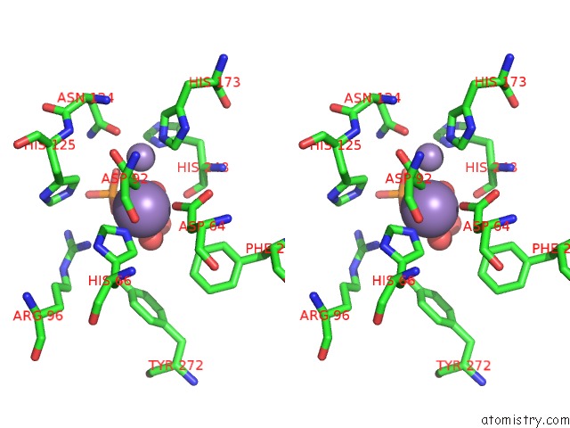
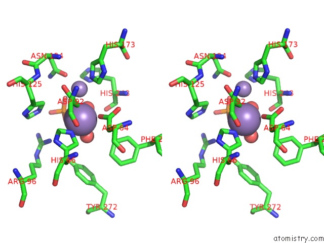
Manganese binding site 3 out of 4 in 4mov

Go back to Manganese Binding Sites List in 4mov
Manganese binding site 3 out of 4 in the 1.45 A Resolution Crystal Structure of Protein Phosphatase 1


Mono view


Stereo pair view

A full contact list of Manganese with other atoms in the Mn binding site number 3 of 1.45 A Resolution Crystal Structure of Protein Phosphatase 1 within 5.0Å range:
probe atom residue distance (Å) B Occ
B:Mn401

b:9.8
occ:1.00
OD1 B:ASN124 2.1 10.4 1.0
O1 B:PO4403 2.2 11.7 0.8
OD2 B:ASP92 2.2 9.3 1.0
NE2 B:HIS173 2.2 9.6 1.0
ND1 B:HIS248 2.2 10.6 1.0
O B:HOH508 2.3 13.6 1.0
CE1 B:HIS248 3.1 11.5 1.0
CG B:ASP92 3.2 9.5 1.0
CG B:ASN124 3.2 10.0 1.0
CE1 B:HIS173 3.2 10.2 1.0
MN B:MN402 3.2 13.6 0.5
CD2 B:HIS173 3.2 9.8 1.0
CG B:HIS248 3.4 10.6 1.0
P B:PO4403 3.4 18.5 0.8
OD1 B:ASP92 3.5 9.4 1.0
ND2 B:ASN124 3.6 11.3 1.0
O3 B:PO4403 3.7 20.3 0.8
CA B:HIS248 3.8 11.0 1.0
CB B:HIS248 3.9 10.0 1.0
OD2 B:ASP64 3.9 13.2 1.0
O2 B:PO4403 4.1 21.7 0.8
O B:HIS248 4.1 20.6 1.0
CD2 B:HIS125 4.2 9.8 1.0
NE2 B:HIS248 4.2 12.6 1.0
ND1 B:HIS173 4.3 8.4 1.0
CG B:HIS173 4.4 8.0 1.0
CB B:ASP92 4.4 8.1 1.0
CD2 B:HIS248 4.4 13.3 1.0
C B:HIS248 4.5 13.6 1.0
CB B:ASN124 4.5 9.7 1.0
N B:ASN124 4.5 8.7 1.0
O4 B:PO4403 4.6 20.1 0.8
O B:LEU205 4.7 12.1 1.0
NE2 B:HIS125 4.8 10.9 1.0
N B:HIS248 4.9 10.1 1.0
CG B:ASP64 4.9 9.3 1.0
NE2 B:HIS66 4.9 10.6 1.0
CA B:ASN124 5.0 9.3 1.0

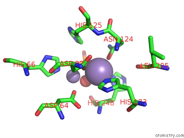
Manganese binding site 4 out of 4 in 4mov

Go back to Manganese Binding Sites List in 4mov
Manganese binding site 4 out of 4 in the 1.45 A Resolution Crystal Structure of Protein Phosphatase 1


Mono view


Stereo pair view

A full contact list of Manganese with other atoms in the Mn binding site number 4 of 1.45 A Resolution Crystal Structure of Protein Phosphatase 1 within 5.0Å range:
probe atom residue distance (Å) B Occ
B:Mn402

b:13.6
occ:0.50
O B:HOH508 1.8 13.6 1.0
NE2 B:HIS66 2.1 10.6 1.0
O3 B:PO4403 2.1 20.3 0.8
O B:HOH685 2.2 28.5 1.0
OD2 B:ASP92 2.3 9.3 1.0
OD2 B:ASP64 2.3 13.2 1.0
CE1 B:HIS66 3.0 9.0 1.0
CD2 B:HIS66 3.1 9.8 1.0
MN B:MN401 3.2 9.8 1.0
P B:PO4403 3.3 18.5 0.8
CG B:ASP92 3.4 9.5 1.0
O1 B:PO4403 3.4 11.7 0.8
CG B:ASP64 3.5 9.3 1.0
CB B:ASP92 3.7 8.1 1.0
O2 B:PO4403 3.9 21.7 0.8
O B:HIS248 4.0 20.6 1.0
CB B:ASP64 4.2 9.0 1.0
ND1 B:HIS66 4.2 8.5 1.0
NH1 B:ARG96 4.2 26.3 1.0
CG B:HIS66 4.2 7.8 1.0
CD2 B:HIS125 4.3 9.8 1.0
CE2 B:PHE267 4.3 10.6 1.0
OH B:TYR272 4.4 34.8 1.0
OD1 B:ASP92 4.5 9.4 1.0
OD1 B:ASP64 4.5 10.9 1.0
O4 B:PO4403 4.5 20.1 0.8
NE2 B:HIS125 4.5 10.9 1.0
NE2 B:HIS173 4.5 9.6 1.0
CE1 B:HIS173 4.6 10.2 1.0
CA B:HIS248 4.7 11.0 1.0
C B:HIS248 4.7 13.6 1.0
ND1 B:HIS248 4.8 10.6 1.0
CZ B:PHE267 4.9 12.5 1.0
OD1 B:ASN124 4.9 10.4 1.0

Reference:

M.S.Choy, M.Hieke, G.S.Kumar, G.R.Lewis, K.R.Gonzalez-Dewhitt, R.P.Kessler, B.J.Stein, M.Hessenberger, A.C.Nairn, W.Peti, R.Page. Understanding the Antagonism of Retinoblastoma Protein Dephosphorylation By Pnuts Provides Insights Into the PP1 Regulatory Code. Proc.Natl.Acad.Sci.Usa V. 111 4097 2014.
ISSN: ISSN 0027-8424
PubMed: 24591642
DOI: 10.1073/PNAS.1317395111
Page generated: Tue Dec 15 04:24:38 2020

Last articles

Zn in 8WB0
Zn in 8WAX
Zn in 8WAU
Zn in 8WAZ
Zn in 8WAY
Zn in 8WAV
Zn in 8WAW
Zn in 8WAT
Zn in 8W7M
Zn in 8WD3
© Copyright 2008-2020 by atomistry.com
Home   |    Site Map   |    Copyright   |    Contact us   |    Privacy