Manganese »
PDB 4lta-4mu3 »
4lul »
Manganese in PDB 4lul: The Crystal Structure of the P132A, Y133D Mutant of Pyrococcus Furiosus Phosphoglucose Isomerase in Complex with Manganese.
Enzymatic activity of The Crystal Structure of the P132A, Y133D Mutant of Pyrococcus Furiosus Phosphoglucose Isomerase in Complex with Manganese.
All present enzymatic activity of The Crystal Structure of the P132A, Y133D Mutant of Pyrococcus Furiosus Phosphoglucose Isomerase in Complex with Manganese.:
5.3.1.9;
5.3.1.9;
Protein crystallography data
The structure of The Crystal Structure of the P132A, Y133D Mutant of Pyrococcus Furiosus Phosphoglucose Isomerase in Complex with Manganese., PDB code: 4lul
was solved by
P.J.Baker,
F.M.Almourfi,
with X-Ray Crystallography technique. A brief refinement statistics is given in the table below:
| Resolution Low / High (Å) | 25.58 / 1.89 |
| Space group | P 21 21 21 |
| Cell size a, b, c (Å), α, β, γ (°) | 41.300, 60.030, 146.670, 90.00, 90.00, 90.00 |
| R / Rfree (%) | 19.6 / 25.3 |
Manganese Binding Sites:
The binding sites of Manganese atom in the The Crystal Structure of the P132A, Y133D Mutant of Pyrococcus Furiosus Phosphoglucose Isomerase in Complex with Manganese.
(pdb code 4lul). This binding sites where shown within
5.0 Angstroms radius around Manganese atom.
In total 2 binding sites of Manganese where determined in the The Crystal Structure of the P132A, Y133D Mutant of Pyrococcus Furiosus Phosphoglucose Isomerase in Complex with Manganese., PDB code: 4lul:
Jump to Manganese binding site number: 1; 2;
In total 2 binding sites of Manganese where determined in the The Crystal Structure of the P132A, Y133D Mutant of Pyrococcus Furiosus Phosphoglucose Isomerase in Complex with Manganese., PDB code: 4lul:
Jump to Manganese binding site number: 1; 2;
Manganese binding site 1 out of 2 in 4lul
Go back to
Manganese binding site 1 out
of 2 in the The Crystal Structure of the P132A, Y133D Mutant of Pyrococcus Furiosus Phosphoglucose Isomerase in Complex with Manganese.
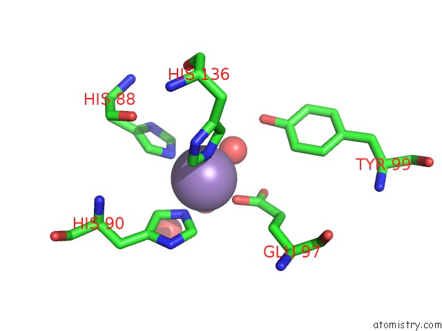
Mono view
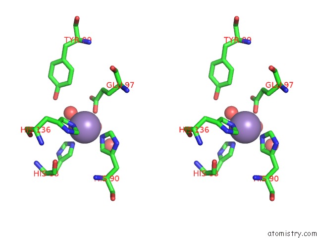
Stereo pair view
Mono view
Stereo pair view
A full contact list of Manganese with other atoms in the Mn binding
site number 1 of The Crystal Structure of the P132A, Y133D Mutant of Pyrococcus Furiosus Phosphoglucose Isomerase in Complex with Manganese. within 5.0Å range:
|
Manganese binding site 2 out of 2 in 4lul
Go back to
Manganese binding site 2 out
of 2 in the The Crystal Structure of the P132A, Y133D Mutant of Pyrococcus Furiosus Phosphoglucose Isomerase in Complex with Manganese.
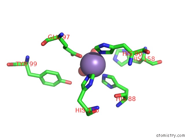
Mono view
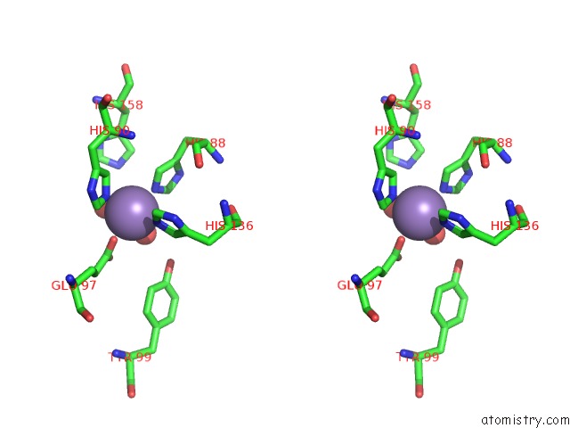
Stereo pair view
Mono view
Stereo pair view
A full contact list of Manganese with other atoms in the Mn binding
site number 2 of The Crystal Structure of the P132A, Y133D Mutant of Pyrococcus Furiosus Phosphoglucose Isomerase in Complex with Manganese. within 5.0Å range:
|
Reference:
P.J.Baker,
F.M.Almourfi,
J.Raedts,
H-J.Joosten,
S.Hendriks,
S.W.M.Kengen,
W.R.Hage,
P.J.Schaap,
S.E.Sedelnikova,
J.Van Der Oost.
Correlated Mutation Analysis As A Tool For Smart Library Design to Improve Protein Performance. To Be Published.
Page generated: Sat Oct 5 20:16:42 2024
Last articles
Zn in 9JYWZn in 9IR4
Zn in 9IR3
Zn in 9GMX
Zn in 9GMW
Zn in 9JEJ
Zn in 9ERF
Zn in 9ERE
Zn in 9EGV
Zn in 9EGW