Manganese »
PDB 4k3v-4lt5 »
4lac »
Manganese in PDB 4lac: Crystal Structure of Protein Phosphatase 2A (PP2A) and PP2A Phosphatase Activator (Ptpa) Complex with Atpgammas
Enzymatic activity of Crystal Structure of Protein Phosphatase 2A (PP2A) and PP2A Phosphatase Activator (Ptpa) Complex with Atpgammas
All present enzymatic activity of Crystal Structure of Protein Phosphatase 2A (PP2A) and PP2A Phosphatase Activator (Ptpa) Complex with Atpgammas:
3.1.3.16; 5.2.1.8;
3.1.3.16; 5.2.1.8;
Protein crystallography data
The structure of Crystal Structure of Protein Phosphatase 2A (PP2A) and PP2A Phosphatase Activator (Ptpa) Complex with Atpgammas, PDB code: 4lac
was solved by
F.Guo,
V.Stanevich,
N.Wlodarchak,
K.A.Satyshur,
Y.Xing,
with X-Ray Crystallography technique. A brief refinement statistics is given in the table below:
| Resolution Low / High (Å) | 50.00 / 2.82 |
| Space group | P 21 21 21 |
| Cell size a, b, c (Å), α, β, γ (°) | 57.070, 100.186, 167.450, 90.00, 90.00, 90.00 |
| R / Rfree (%) | 18.8 / 24.2 |
Manganese Binding Sites:
The binding sites of Manganese atom in the Crystal Structure of Protein Phosphatase 2A (PP2A) and PP2A Phosphatase Activator (Ptpa) Complex with Atpgammas
(pdb code 4lac). This binding sites where shown within
5.0 Angstroms radius around Manganese atom.
In total 2 binding sites of Manganese where determined in the Crystal Structure of Protein Phosphatase 2A (PP2A) and PP2A Phosphatase Activator (Ptpa) Complex with Atpgammas, PDB code: 4lac:
Jump to Manganese binding site number: 1; 2;
In total 2 binding sites of Manganese where determined in the Crystal Structure of Protein Phosphatase 2A (PP2A) and PP2A Phosphatase Activator (Ptpa) Complex with Atpgammas, PDB code: 4lac:
Jump to Manganese binding site number: 1; 2;
Manganese binding site 1 out of 2 in 4lac
Go back to
Manganese binding site 1 out
of 2 in the Crystal Structure of Protein Phosphatase 2A (PP2A) and PP2A Phosphatase Activator (Ptpa) Complex with Atpgammas
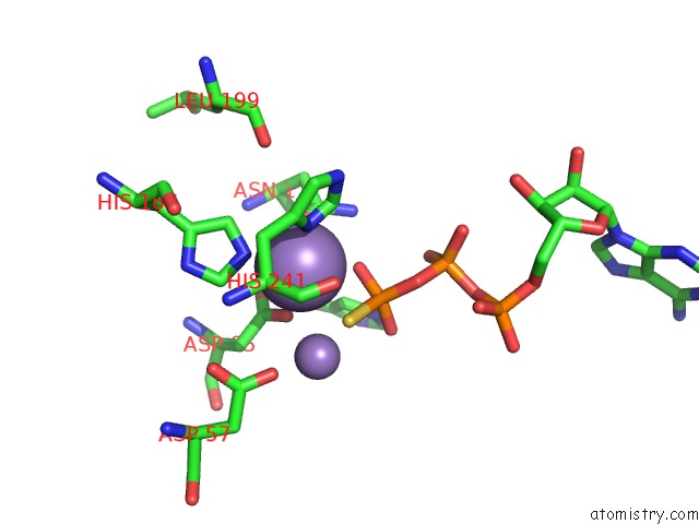
Mono view
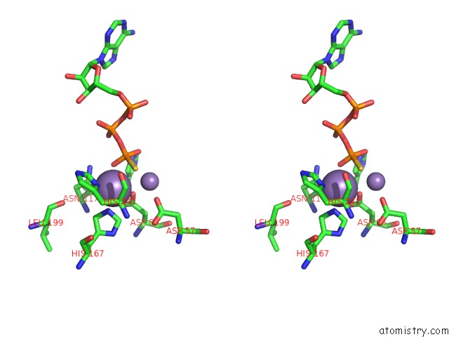
Stereo pair view
Mono view
Stereo pair view
A full contact list of Manganese with other atoms in the Mn binding
site number 1 of Crystal Structure of Protein Phosphatase 2A (PP2A) and PP2A Phosphatase Activator (Ptpa) Complex with Atpgammas within 5.0Å range:
|
Manganese binding site 2 out of 2 in 4lac
Go back to
Manganese binding site 2 out
of 2 in the Crystal Structure of Protein Phosphatase 2A (PP2A) and PP2A Phosphatase Activator (Ptpa) Complex with Atpgammas
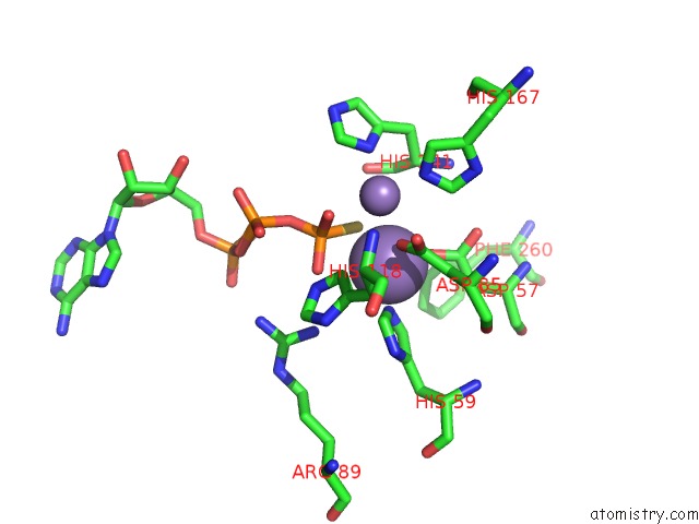
Mono view
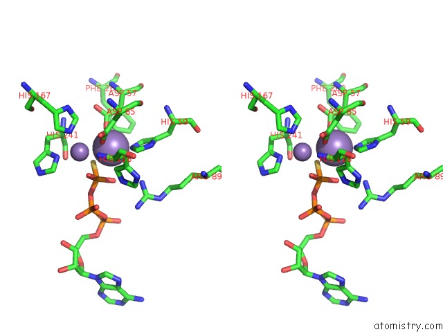
Stereo pair view
Mono view
Stereo pair view
A full contact list of Manganese with other atoms in the Mn binding
site number 2 of Crystal Structure of Protein Phosphatase 2A (PP2A) and PP2A Phosphatase Activator (Ptpa) Complex with Atpgammas within 5.0Å range:
|
Reference:
F.Guo,
V.Stanevich,
N.Wlodarchak,
R.Sengupta,
L.Jiang,
K.A.Satyshur,
Y.Xing.
Structural Basis of PP2A Activation By Ptpa, An Atp-Dependent Activation Chaperone. Cell Res. V. 24 190 2014.
ISSN: ISSN 1001-0602
PubMed: 24100351
DOI: 10.1038/CR.2013.138
Page generated: Sat Oct 5 20:06:35 2024
ISSN: ISSN 1001-0602
PubMed: 24100351
DOI: 10.1038/CR.2013.138
Last articles
Cl in 7UP4Cl in 7UOS
Cl in 7UPI
Cl in 7UMO
Cl in 7UOD
Cl in 7UN0
Cl in 7UOQ
Cl in 7UOC
Cl in 7UNO
Cl in 7UNN