Manganese »
PDB 4k3v-4lt5 »
4kz2 »
Manganese in PDB 4kz2: Crystal Structure of PHI29 Prna 3WJ Core
Protein crystallography data
The structure of Crystal Structure of PHI29 Prna 3WJ Core, PDB code: 4kz2
was solved by
H.Zhang,
J.A.Endrizzi,
Y.Shu,
F.Haque,
C.Sauter,
P.Guo,
Y.-I.Chi,
with X-Ray Crystallography technique. A brief refinement statistics is given in the table below:
| Resolution Low / High (Å) | 30.00 / 3.05 |
| Space group | I 4 |
| Cell size a, b, c (Å), α, β, γ (°) | 125.012, 125.012, 26.989, 90.00, 90.00, 90.00 |
| R / Rfree (%) | 19.7 / 19.7 |
Manganese Binding Sites:
The binding sites of Manganese atom in the Crystal Structure of PHI29 Prna 3WJ Core
(pdb code 4kz2). This binding sites where shown within
5.0 Angstroms radius around Manganese atom.
In total 3 binding sites of Manganese where determined in the Crystal Structure of PHI29 Prna 3WJ Core, PDB code: 4kz2:
Jump to Manganese binding site number: 1; 2; 3;
In total 3 binding sites of Manganese where determined in the Crystal Structure of PHI29 Prna 3WJ Core, PDB code: 4kz2:
Jump to Manganese binding site number: 1; 2; 3;
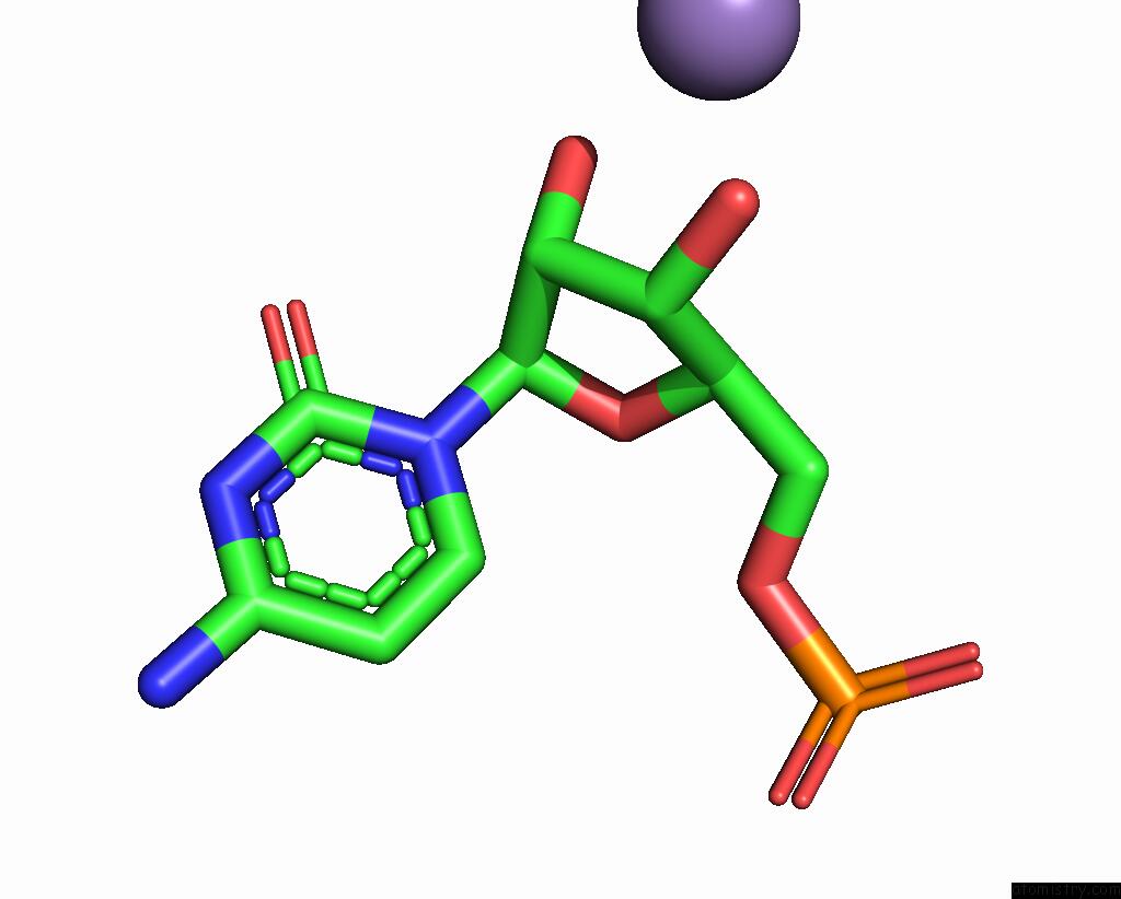
Manganese binding site 1 out of 3 in 4kz2
Go back to
Manganese binding site 1 out
of 3 in the Crystal Structure of PHI29 Prna 3WJ Core

Mono view
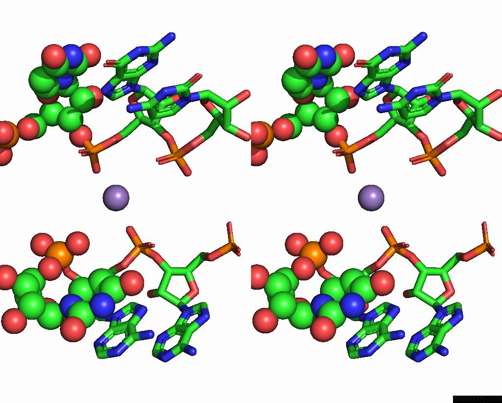
Stereo pair view
Mono view
Stereo pair view
A full contact list of Manganese with other atoms in the Mn binding
site number 1 of Crystal Structure of PHI29 Prna 3WJ Core within 5.0Å range:
|
Manganese binding site 2 out of 3 in 4kz2
Go back to
Manganese binding site 2 out
of 3 in the Crystal Structure of PHI29 Prna 3WJ Core
Mono view

Stereo pair view
Mono view
Stereo pair view
A full contact list of Manganese with other atoms in the Mn binding
site number 2 of Crystal Structure of PHI29 Prna 3WJ Core within 5.0Å range:
|
Manganese binding site 3 out of 3 in 4kz2
Go back to
Manganese binding site 3 out
of 3 in the Crystal Structure of PHI29 Prna 3WJ Core

Mono view

Stereo pair view
Mono view
Stereo pair view
A full contact list of Manganese with other atoms in the Mn binding
site number 3 of Crystal Structure of PHI29 Prna 3WJ Core within 5.0Å range:
|
Reference:
H.Zhang,
J.A.Endrizzi,
Y.Shu,
F.Haque,
C.Sauter,
L.S.Shlyakhtenko,
Y.Lyubchenko,
P.Guo,
Y.I.Chi.
Crystal Structure of 3WJ Core Revealing Divalent Ion-Promoted Thermostability and Assembly of the PHI29 Hexameric Motor Prna. Rna V. 19 1226 2013.
ISSN: ISSN 1355-8382
PubMed: 23884902
DOI: 10.1261/RNA.037077.112
Page generated: Sat Oct 5 20:05:34 2024
ISSN: ISSN 1355-8382
PubMed: 23884902
DOI: 10.1261/RNA.037077.112
Last articles
Zn in 9MJ5Zn in 9HNW
Zn in 9G0L
Zn in 9FNE
Zn in 9DZN
Zn in 9E0I
Zn in 9D32
Zn in 9DAK
Zn in 8ZXC
Zn in 8ZUF