Manganese »
PDB 4ima-4k28 »
4k28 »
Manganese in PDB 4k28: 2.15 Angstrom Resolution Crystal Structure of A Shikimate Dehydrogenase Family Protein From Pseudomonas Putida KT2440 in Complex with Nad+
Protein crystallography data
The structure of 2.15 Angstrom Resolution Crystal Structure of A Shikimate Dehydrogenase Family Protein From Pseudomonas Putida KT2440 in Complex with Nad+, PDB code: 4k28
was solved by
C.Garcia,
J.Peek,
P.Petit,
D.Christendat,
with X-Ray Crystallography technique. A brief refinement statistics is given in the table below:
| Resolution Low / High (Å) | 19.22 / 2.15 |
| Space group | P 21 21 21 |
| Cell size a, b, c (Å), α, β, γ (°) | 44.437, 76.688, 153.333, 90.00, 90.00, 90.00 |
| R / Rfree (%) | 17.9 / 23.8 |
Other elements in 4k28:
The structure of 2.15 Angstrom Resolution Crystal Structure of A Shikimate Dehydrogenase Family Protein From Pseudomonas Putida KT2440 in Complex with Nad+ also contains other interesting chemical elements:
| Sodium | (Na) | 1 atom |
Manganese Binding Sites:
The binding sites of Manganese atom in the 2.15 Angstrom Resolution Crystal Structure of A Shikimate Dehydrogenase Family Protein From Pseudomonas Putida KT2440 in Complex with Nad+
(pdb code 4k28). This binding sites where shown within
5.0 Angstroms radius around Manganese atom.
In total 6 binding sites of Manganese where determined in the 2.15 Angstrom Resolution Crystal Structure of A Shikimate Dehydrogenase Family Protein From Pseudomonas Putida KT2440 in Complex with Nad+, PDB code: 4k28:
Jump to Manganese binding site number: 1; 2; 3; 4; 5; 6;
In total 6 binding sites of Manganese where determined in the 2.15 Angstrom Resolution Crystal Structure of A Shikimate Dehydrogenase Family Protein From Pseudomonas Putida KT2440 in Complex with Nad+, PDB code: 4k28:
Jump to Manganese binding site number: 1; 2; 3; 4; 5; 6;
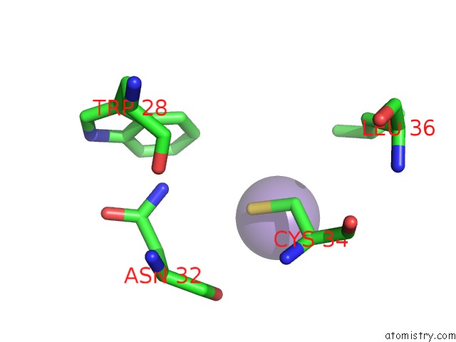
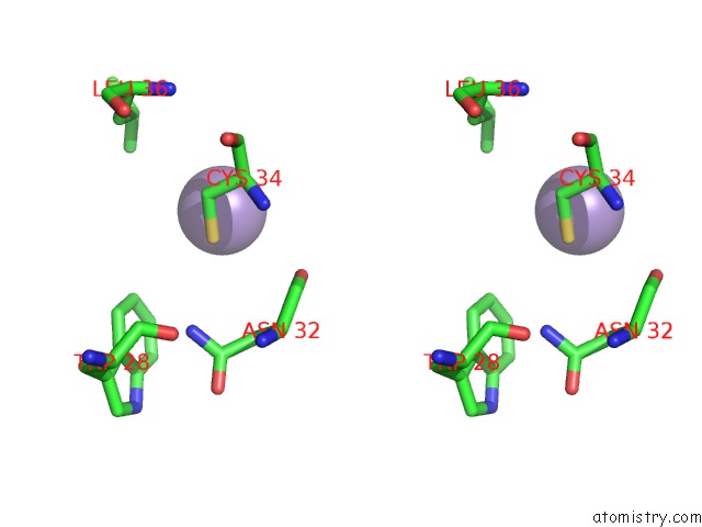
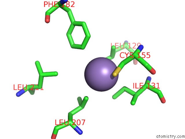
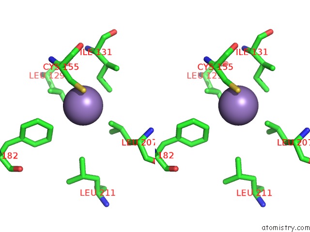
Manganese binding site 1 out of 6 in 4k28
Go back to
Manganese binding site 1 out
of 6 in the 2.15 Angstrom Resolution Crystal Structure of A Shikimate Dehydrogenase Family Protein From Pseudomonas Putida KT2440 in Complex with Nad+
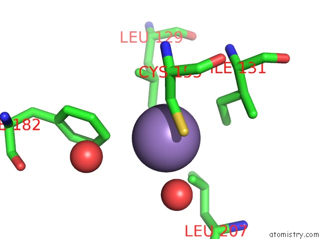
Mono view
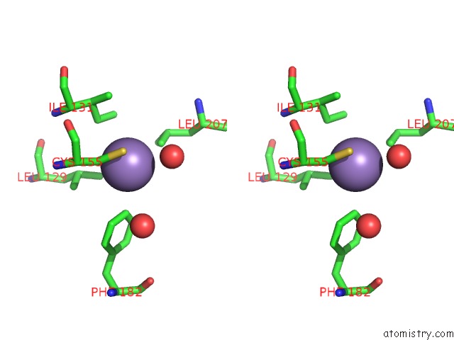
Stereo pair view
Mono view
Stereo pair view
A full contact list of Manganese with other atoms in the Mn binding
site number 1 of 2.15 Angstrom Resolution Crystal Structure of A Shikimate Dehydrogenase Family Protein From Pseudomonas Putida KT2440 in Complex with Nad+ within 5.0Å range:
|
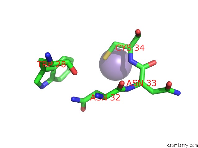
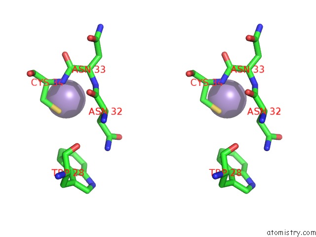
Manganese binding site 2 out of 6 in 4k28
Go back to
Manganese binding site 2 out
of 6 in the 2.15 Angstrom Resolution Crystal Structure of A Shikimate Dehydrogenase Family Protein From Pseudomonas Putida KT2440 in Complex with Nad+
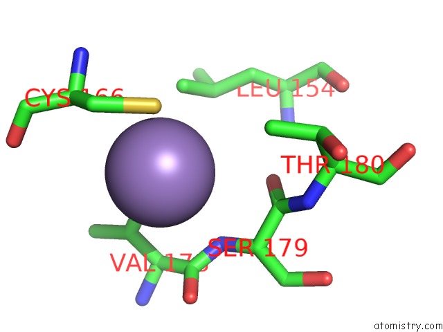
Mono view
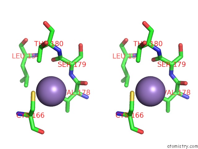
Stereo pair view
Mono view
Stereo pair view
A full contact list of Manganese with other atoms in the Mn binding
site number 2 of 2.15 Angstrom Resolution Crystal Structure of A Shikimate Dehydrogenase Family Protein From Pseudomonas Putida KT2440 in Complex with Nad+ within 5.0Å range:
|
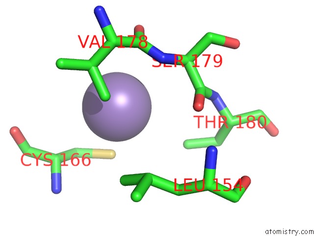
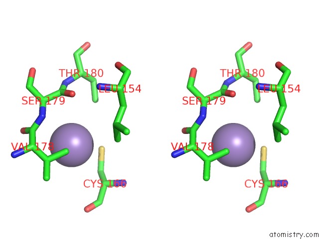
Manganese binding site 3 out of 6 in 4k28
Go back to
Manganese binding site 3 out
of 6 in the 2.15 Angstrom Resolution Crystal Structure of A Shikimate Dehydrogenase Family Protein From Pseudomonas Putida KT2440 in Complex with Nad+
Mono view
Stereo pair view
Mono view
Stereo pair view
A full contact list of Manganese with other atoms in the Mn binding
site number 3 of 2.15 Angstrom Resolution Crystal Structure of A Shikimate Dehydrogenase Family Protein From Pseudomonas Putida KT2440 in Complex with Nad+ within 5.0Å range:
|
Manganese binding site 4 out of 6 in 4k28
Go back to
Manganese binding site 4 out
of 6 in the 2.15 Angstrom Resolution Crystal Structure of A Shikimate Dehydrogenase Family Protein From Pseudomonas Putida KT2440 in Complex with Nad+
Mono view
Stereo pair view
Mono view
Stereo pair view
A full contact list of Manganese with other atoms in the Mn binding
site number 4 of 2.15 Angstrom Resolution Crystal Structure of A Shikimate Dehydrogenase Family Protein From Pseudomonas Putida KT2440 in Complex with Nad+ within 5.0Å range:
|
Manganese binding site 5 out of 6 in 4k28
Go back to
Manganese binding site 5 out
of 6 in the 2.15 Angstrom Resolution Crystal Structure of A Shikimate Dehydrogenase Family Protein From Pseudomonas Putida KT2440 in Complex with Nad+
Mono view
Stereo pair view
Mono view
Stereo pair view
A full contact list of Manganese with other atoms in the Mn binding
site number 5 of 2.15 Angstrom Resolution Crystal Structure of A Shikimate Dehydrogenase Family Protein From Pseudomonas Putida KT2440 in Complex with Nad+ within 5.0Å range:
|
Manganese binding site 6 out of 6 in 4k28
Go back to
Manganese binding site 6 out
of 6 in the 2.15 Angstrom Resolution Crystal Structure of A Shikimate Dehydrogenase Family Protein From Pseudomonas Putida KT2440 in Complex with Nad+
Mono view
Stereo pair view
Mono view
Stereo pair view
A full contact list of Manganese with other atoms in the Mn binding
site number 6 of 2.15 Angstrom Resolution Crystal Structure of A Shikimate Dehydrogenase Family Protein From Pseudomonas Putida KT2440 in Complex with Nad+ within 5.0Å range:
|
Reference:
J.Peek,
C.Garcia,
J.Lee,
D.Christendat.
Insights Into the Function of RIFI2: Structural and Biochemical Investigation of A New Shikimate Dehydrogenase Family Protein. Biochim.Biophys.Acta V.1834 516 2013.
ISSN: ISSN 0006-3002
PubMed: 23142411
DOI: 10.1016/J.BBAPAP.2012.10.016
Page generated: Sat Oct 5 20:00:25 2024
ISSN: ISSN 0006-3002
PubMed: 23142411
DOI: 10.1016/J.BBAPAP.2012.10.016
Last articles
Zn in 9J0NZn in 9J0O
Zn in 9J0P
Zn in 9FJX
Zn in 9EKB
Zn in 9C0F
Zn in 9CAH
Zn in 9CH0
Zn in 9CH3
Zn in 9CH1