Manganese »
PDB 4ima-4k28 »
4j0n »
Manganese in PDB 4j0n: Crystal Structure of A Manganese Dependent Isatin Hydrolase
Protein crystallography data
The structure of Crystal Structure of A Manganese Dependent Isatin Hydrolase, PDB code: 4j0n
was solved by
K.Bjerregaard-Andersen,
T.Sommer,
J.K.Jensen,
B.Jochimsen,
M.Etzerodt,
J.P.Morth,
with X-Ray Crystallography technique. A brief refinement statistics is given in the table below:
| Resolution Low / High (Å) | 43.59 / 2.25 |
| Space group | P 32 |
| Cell size a, b, c (Å), α, β, γ (°) | 51.940, 51.940, 176.690, 90.00, 90.00, 120.00 |
| R / Rfree (%) | 17.1 / 22.3 |
Other elements in 4j0n:
The structure of Crystal Structure of A Manganese Dependent Isatin Hydrolase also contains other interesting chemical elements:
| Calcium | (Ca) | 1 atom |
| Sodium | (Na) | 3 atoms |
Manganese Binding Sites:
The binding sites of Manganese atom in the Crystal Structure of A Manganese Dependent Isatin Hydrolase
(pdb code 4j0n). This binding sites where shown within
5.0 Angstroms radius around Manganese atom.
In total 2 binding sites of Manganese where determined in the Crystal Structure of A Manganese Dependent Isatin Hydrolase, PDB code: 4j0n:
Jump to Manganese binding site number: 1; 2;
In total 2 binding sites of Manganese where determined in the Crystal Structure of A Manganese Dependent Isatin Hydrolase, PDB code: 4j0n:
Jump to Manganese binding site number: 1; 2;
Manganese binding site 1 out of 2 in 4j0n
Go back to
Manganese binding site 1 out
of 2 in the Crystal Structure of A Manganese Dependent Isatin Hydrolase
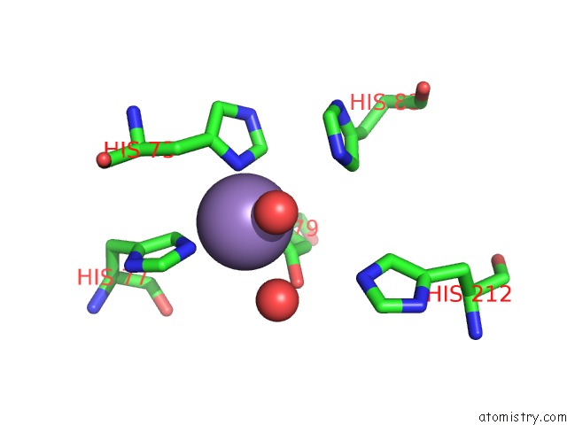
Mono view
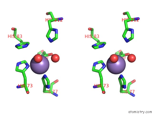
Stereo pair view
Mono view
Stereo pair view
A full contact list of Manganese with other atoms in the Mn binding
site number 1 of Crystal Structure of A Manganese Dependent Isatin Hydrolase within 5.0Å range:
|
Manganese binding site 2 out of 2 in 4j0n
Go back to
Manganese binding site 2 out
of 2 in the Crystal Structure of A Manganese Dependent Isatin Hydrolase
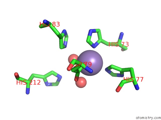
Mono view
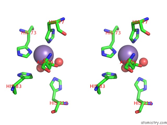
Stereo pair view
Mono view
Stereo pair view
A full contact list of Manganese with other atoms in the Mn binding
site number 2 of Crystal Structure of A Manganese Dependent Isatin Hydrolase within 5.0Å range:
|
Reference:
K.Bjerregaard-Andersen,
T.Sommer,
J.K.Jensen,
B.Jochimsen,
M.Etzerodt,
J.P.Morth.
A Proton Wire and Water Channel Revealed in the Crystal Structure of Isatin Hydrolase. J.Biol.Chem. V. 289 21351 2014.
ISSN: ISSN 0021-9258
PubMed: 24917679
DOI: 10.1074/JBC.M114.568824
Page generated: Sat Oct 5 19:56:28 2024
ISSN: ISSN 0021-9258
PubMed: 24917679
DOI: 10.1074/JBC.M114.568824
Last articles
Zn in 9MJ5Zn in 9HNW
Zn in 9G0L
Zn in 9FNE
Zn in 9DZN
Zn in 9E0I
Zn in 9D32
Zn in 9DAK
Zn in 8ZXC
Zn in 8ZUF