Manganese »
PDB 4ima-4k28 »
4ixr »
Manganese in PDB 4ixr: Rt Fs X-Ray Diffraction of Photosystem II, First Illuminated State
Enzymatic activity of Rt Fs X-Ray Diffraction of Photosystem II, First Illuminated State
All present enzymatic activity of Rt Fs X-Ray Diffraction of Photosystem II, First Illuminated State:
1.10.3.9;
1.10.3.9;
Protein crystallography data
The structure of Rt Fs X-Ray Diffraction of Photosystem II, First Illuminated State, PDB code: 4ixr
was solved by
J.Kern,
R.Alonso-Mori,
R.Tran,
J.Hattne,
R.J.Gildea,
N.Echols,
C.Gloeckner,
J.Hellmich,
H.Laksmono,
R.G.Sierra,
B.Lassalle-Kaiser,
S.Koroidov,
A.Lampe,
G.Han,
S.Gul,
D.Difiore,
D.Milathianaki,
A.R.Fry,
A.Miahnahri,
D.W.Schafer,
M.Messerschmidt,
M.M.Seibert,
J.E.Koglin,
D.Sokaras,
T.-C.Weng,
J.Sellberg,
M.J.Latimer,
R.W.Grosse-Kunstleve,
P.H.Zwart,
W.E.White,
P.Glatzel,
P.D.Adams,
M.J.Bogan,
G.J.Williams,
S.Boutet,
J.Messinger,
A.Zouni,
N.K.Sauter,
V.K.Yachandra,
U.Bergmann,
J.Yano,
with X-Ray Crystallography technique. A brief refinement statistics is given in the table below:
| Resolution Low / High (Å) | 82.97 / 5.90 |
| Space group | P 21 21 21 |
| Cell size a, b, c (Å), α, β, γ (°) | 131.976, 227.568, 306.994, 90.00, 90.00, 90.00 |
| R / Rfree (%) | 28.5 / 31.3 |
Other elements in 4ixr:
The structure of Rt Fs X-Ray Diffraction of Photosystem II, First Illuminated State also contains other interesting chemical elements:
| Magnesium | (Mg) | 70 atoms |
| Iron | (Fe) | 6 atoms |
| Chlorine | (Cl) | 2 atoms |
| Calcium | (Ca) | 6 atoms |
Manganese Binding Sites:
The binding sites of Manganese atom in the Rt Fs X-Ray Diffraction of Photosystem II, First Illuminated State
(pdb code 4ixr). This binding sites where shown within
5.0 Angstroms radius around Manganese atom.
In total 8 binding sites of Manganese where determined in the Rt Fs X-Ray Diffraction of Photosystem II, First Illuminated State, PDB code: 4ixr:
Jump to Manganese binding site number: 1; 2; 3; 4; 5; 6; 7; 8;
In total 8 binding sites of Manganese where determined in the Rt Fs X-Ray Diffraction of Photosystem II, First Illuminated State, PDB code: 4ixr:
Jump to Manganese binding site number: 1; 2; 3; 4; 5; 6; 7; 8;
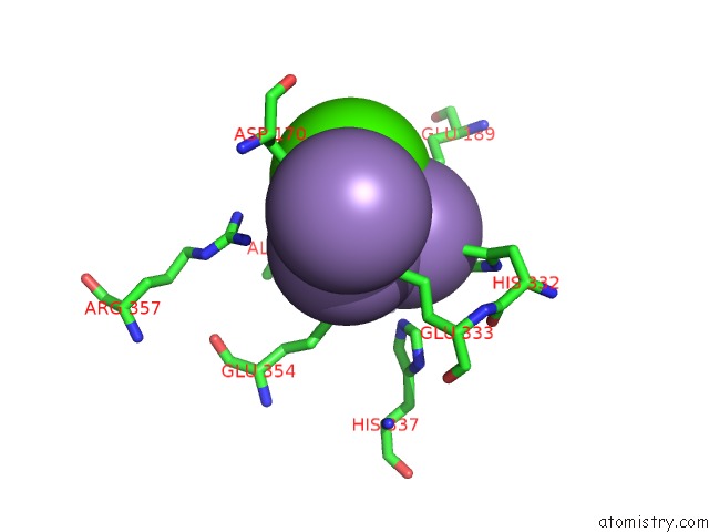
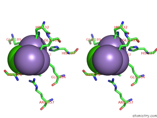
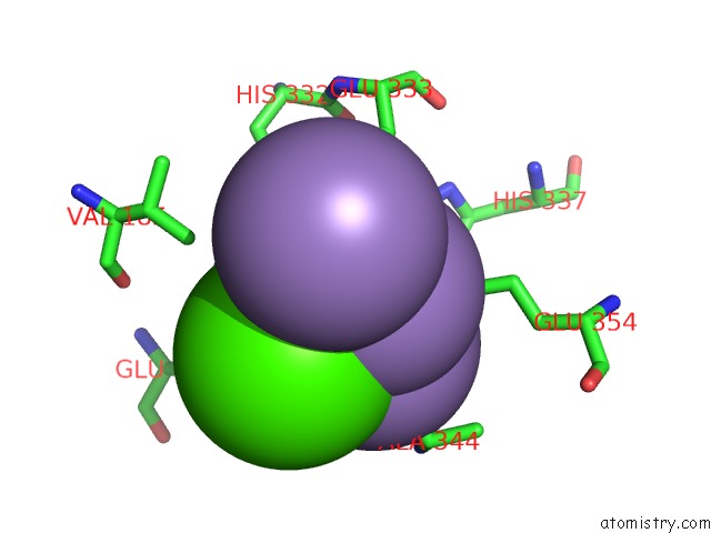
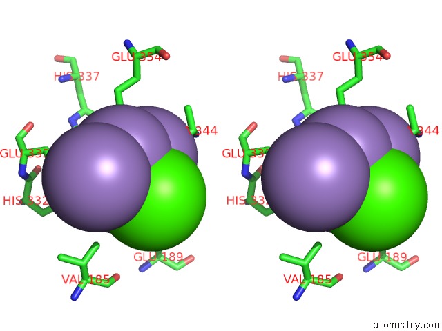
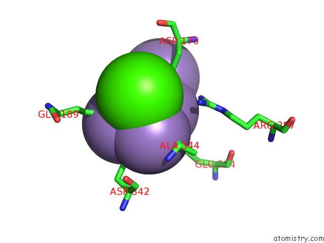
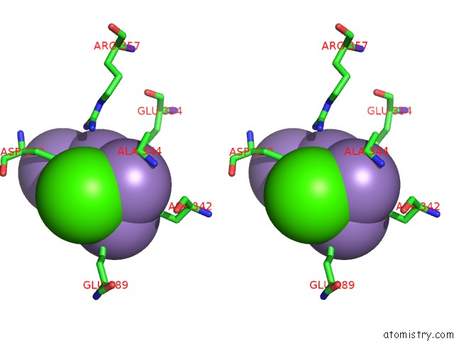
Manganese binding site 1 out of 8 in 4ixr
Go back to
Manganese binding site 1 out
of 8 in the Rt Fs X-Ray Diffraction of Photosystem II, First Illuminated State
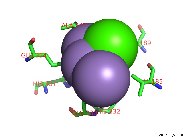
Mono view
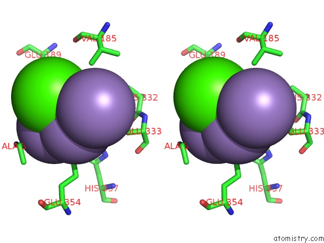
Stereo pair view
Mono view
Stereo pair view
A full contact list of Manganese with other atoms in the Mn binding
site number 1 of Rt Fs X-Ray Diffraction of Photosystem II, First Illuminated State within 5.0Å range:
|
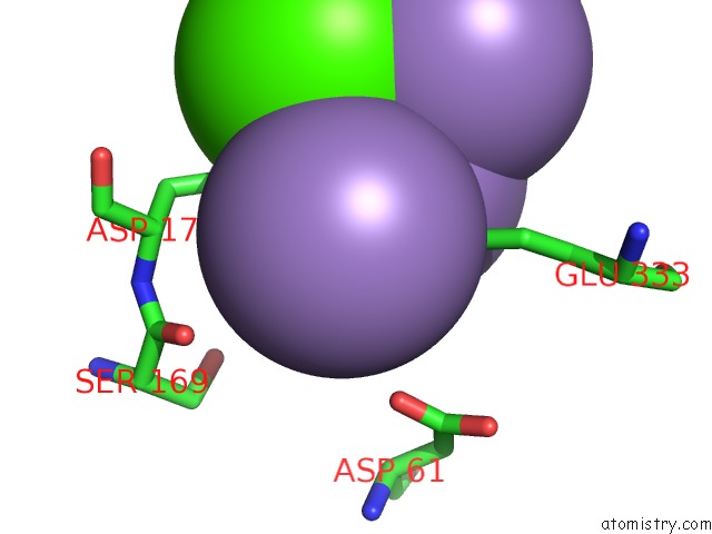
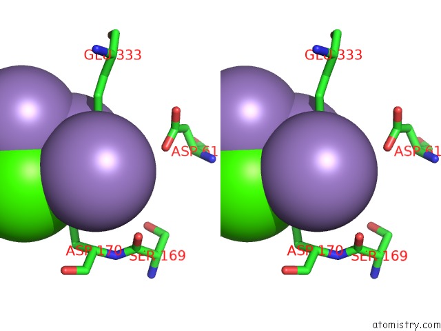
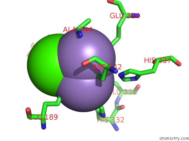
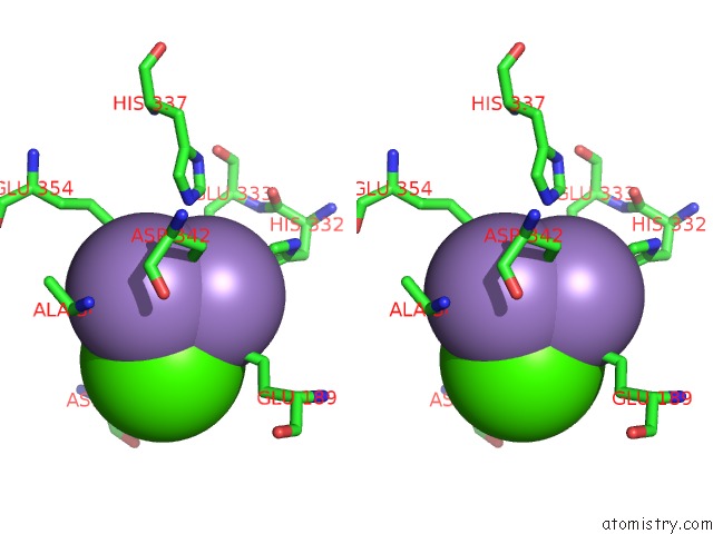
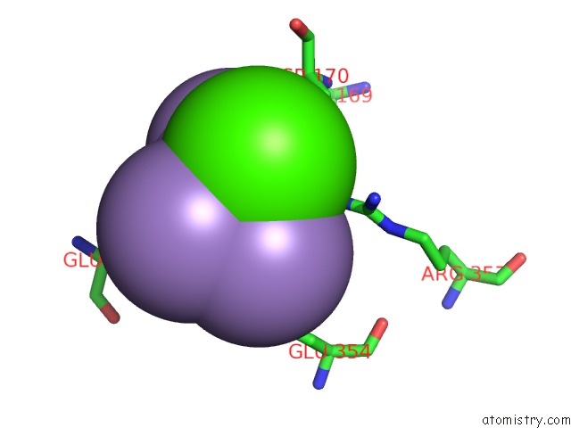
Manganese binding site 2 out of 8 in 4ixr
Go back to
Manganese binding site 2 out
of 8 in the Rt Fs X-Ray Diffraction of Photosystem II, First Illuminated State
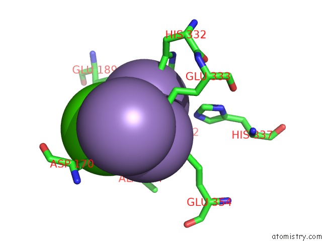
Mono view
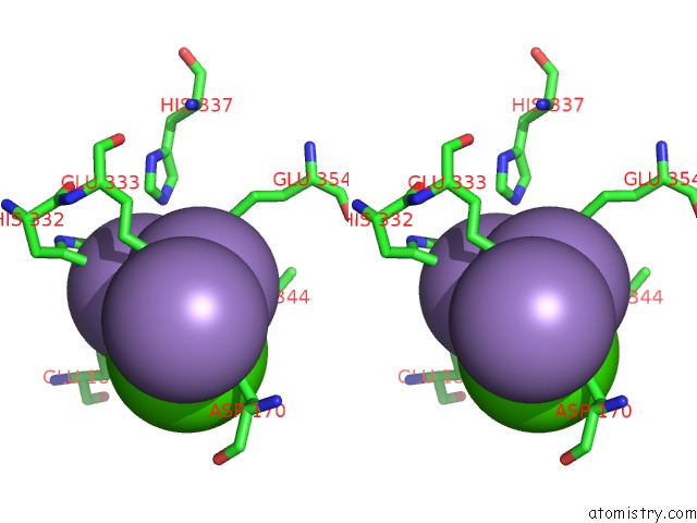
Stereo pair view
Mono view
Stereo pair view
A full contact list of Manganese with other atoms in the Mn binding
site number 2 of Rt Fs X-Ray Diffraction of Photosystem II, First Illuminated State within 5.0Å range:
|
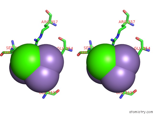
Manganese binding site 3 out of 8 in 4ixr
Go back to
Manganese binding site 3 out
of 8 in the Rt Fs X-Ray Diffraction of Photosystem II, First Illuminated State
Mono view
Stereo pair view
Mono view
Stereo pair view
A full contact list of Manganese with other atoms in the Mn binding
site number 3 of Rt Fs X-Ray Diffraction of Photosystem II, First Illuminated State within 5.0Å range:
|
Manganese binding site 4 out of 8 in 4ixr
Go back to
Manganese binding site 4 out
of 8 in the Rt Fs X-Ray Diffraction of Photosystem II, First Illuminated State
Mono view
Stereo pair view
Mono view
Stereo pair view
A full contact list of Manganese with other atoms in the Mn binding
site number 4 of Rt Fs X-Ray Diffraction of Photosystem II, First Illuminated State within 5.0Å range:
|
Manganese binding site 5 out of 8 in 4ixr
Go back to
Manganese binding site 5 out
of 8 in the Rt Fs X-Ray Diffraction of Photosystem II, First Illuminated State
Mono view
Stereo pair view
Mono view
Stereo pair view
A full contact list of Manganese with other atoms in the Mn binding
site number 5 of Rt Fs X-Ray Diffraction of Photosystem II, First Illuminated State within 5.0Å range:
|
Manganese binding site 6 out of 8 in 4ixr
Go back to
Manganese binding site 6 out
of 8 in the Rt Fs X-Ray Diffraction of Photosystem II, First Illuminated State
Mono view
Stereo pair view
Mono view
Stereo pair view
A full contact list of Manganese with other atoms in the Mn binding
site number 6 of Rt Fs X-Ray Diffraction of Photosystem II, First Illuminated State within 5.0Å range:
|
Manganese binding site 7 out of 8 in 4ixr
Go back to
Manganese binding site 7 out
of 8 in the Rt Fs X-Ray Diffraction of Photosystem II, First Illuminated State
Mono view
Stereo pair view
Mono view
Stereo pair view
A full contact list of Manganese with other atoms in the Mn binding
site number 7 of Rt Fs X-Ray Diffraction of Photosystem II, First Illuminated State within 5.0Å range:
|
Manganese binding site 8 out of 8 in 4ixr
Go back to
Manganese binding site 8 out
of 8 in the Rt Fs X-Ray Diffraction of Photosystem II, First Illuminated State
Mono view
Stereo pair view
Mono view
Stereo pair view
A full contact list of Manganese with other atoms in the Mn binding
site number 8 of Rt Fs X-Ray Diffraction of Photosystem II, First Illuminated State within 5.0Å range:
|
Reference:
J.Kern,
R.Alonso-Mori,
R.Tran,
J.Hattne,
R.J.Gildea,
N.Echols,
C.Glockner,
J.Hellmich,
H.Laksmono,
R.G.Sierra,
B.Lassalle-Kaiser,
S.Koroidov,
A.Lampe,
G.Han,
S.Gul,
D.Difiore,
D.Milathianaki,
A.R.Fry,
A.Miahnahri,
D.W.Schafer,
M.Messerschmidt,
M.M.Seibert,
J.E.Koglin,
D.Sokaras,
T.C.Weng,
J.Sellberg,
M.J.Latimer,
R.W.Grosse-Kunstleve,
P.H.Zwart,
W.E.White,
P.Glatzel,
P.D.Adams,
M.J.Bogan,
G.J.Williams,
S.Boutet,
J.Messinger,
A.Zouni,
N.K.Sauter,
V.K.Yachandra,
U.Bergmann,
J.Yano.
Simultaneous Femtosecond X-Ray Spectroscopy and Diffraction of Photosystem II at Room Temperature. Science V. 340 491 2013.
ISSN: ISSN 0036-8075
PubMed: 23413188
DOI: 10.1126/SCIENCE.1234273
Page generated: Sat Oct 5 19:55:43 2024
ISSN: ISSN 0036-8075
PubMed: 23413188
DOI: 10.1126/SCIENCE.1234273
Last articles
Zn in 9MJ5Zn in 9HNW
Zn in 9G0L
Zn in 9FNE
Zn in 9DZN
Zn in 9E0I
Zn in 9D32
Zn in 9DAK
Zn in 8ZXC
Zn in 8ZUF