Manganese »
PDB 4ima-4k28 »
4irm »
Manganese in PDB 4irm: Crystal Structure of Mntc R116A Mutant Exhibits Flexibility in the C- Terminal Domain
Protein crystallography data
The structure of Crystal Structure of Mntc R116A Mutant Exhibits Flexibility in the C- Terminal Domain, PDB code: 4irm
was solved by
M.Kanteev,
N.Adir,
with X-Ray Crystallography technique. A brief refinement statistics is given in the table below:
| Resolution Low / High (Å) | 24.50 / 3.50 |
| Space group | P 31 2 1 |
| Cell size a, b, c (Å), α, β, γ (°) | 128.400, 128.400, 90.510, 90.00, 90.00, 120.00 |
| R / Rfree (%) | 29.4 / 30.5 |
Manganese Binding Sites:
The binding sites of Manganese atom in the Crystal Structure of Mntc R116A Mutant Exhibits Flexibility in the C- Terminal Domain
(pdb code 4irm). This binding sites where shown within
5.0 Angstroms radius around Manganese atom.
In total 3 binding sites of Manganese where determined in the Crystal Structure of Mntc R116A Mutant Exhibits Flexibility in the C- Terminal Domain, PDB code: 4irm:
Jump to Manganese binding site number: 1; 2; 3;
In total 3 binding sites of Manganese where determined in the Crystal Structure of Mntc R116A Mutant Exhibits Flexibility in the C- Terminal Domain, PDB code: 4irm:
Jump to Manganese binding site number: 1; 2; 3;
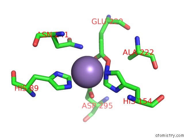
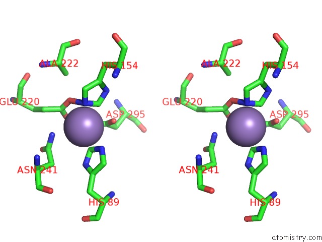
Manganese binding site 1 out of 3 in 4irm
Go back to
Manganese binding site 1 out
of 3 in the Crystal Structure of Mntc R116A Mutant Exhibits Flexibility in the C- Terminal Domain
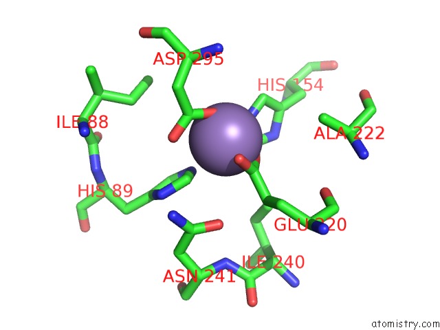
Mono view
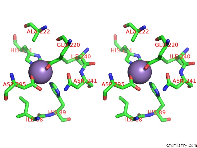
Stereo pair view
Mono view
Stereo pair view
A full contact list of Manganese with other atoms in the Mn binding
site number 1 of Crystal Structure of Mntc R116A Mutant Exhibits Flexibility in the C- Terminal Domain within 5.0Å range:
|
Manganese binding site 2 out of 3 in 4irm
Go back to
Manganese binding site 2 out
of 3 in the Crystal Structure of Mntc R116A Mutant Exhibits Flexibility in the C- Terminal Domain
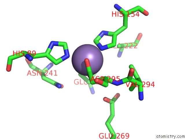
Mono view
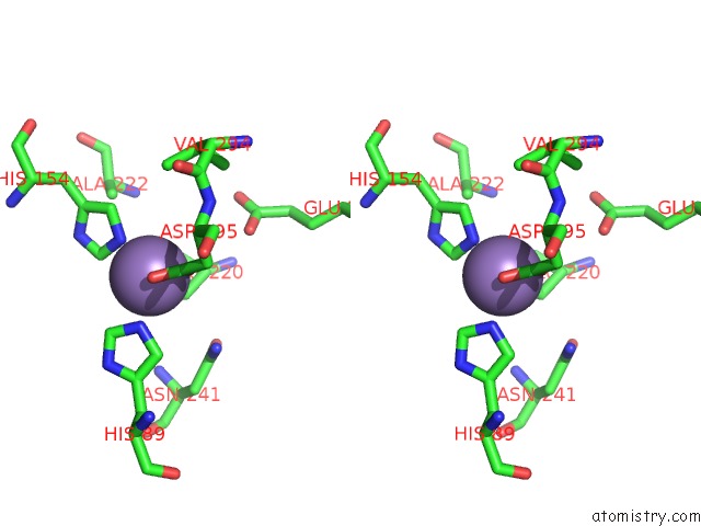
Stereo pair view
Mono view
Stereo pair view
A full contact list of Manganese with other atoms in the Mn binding
site number 2 of Crystal Structure of Mntc R116A Mutant Exhibits Flexibility in the C- Terminal Domain within 5.0Å range:
|
Manganese binding site 3 out of 3 in 4irm
Go back to
Manganese binding site 3 out
of 3 in the Crystal Structure of Mntc R116A Mutant Exhibits Flexibility in the C- Terminal Domain
Mono view
Stereo pair view
Mono view
Stereo pair view
A full contact list of Manganese with other atoms in the Mn binding
site number 3 of Crystal Structure of Mntc R116A Mutant Exhibits Flexibility in the C- Terminal Domain within 5.0Å range:
|
Reference:
M.Kanteev,
N.Adir.
Arginine 116 Stabilizes the Entrance to the Metal Ion-Binding Site of the Mntc Protein. Acta Crystallogr.,Sect.F V. 69 237 2013.
ISSN: ESSN 1744-3091
PubMed: 23519795
DOI: 10.1107/S174430911300153X
Page generated: Sat Oct 5 19:51:56 2024
ISSN: ESSN 1744-3091
PubMed: 23519795
DOI: 10.1107/S174430911300153X
Last articles
Ca in 3AIBCa in 3AII
Ca in 3AIG
Ca in 3AI7
Ca in 3AHW
Ca in 3AFG
Ca in 3AGV
Ca in 3AGO
Ca in 3AGP
Ca in 3ACO