Manganese »
PDB 4gwc-4ilk »
4ie2 »
Manganese in PDB 4ie2: Crystal Structure of Human Arginase-2 Complexed with Inhibitor 1H
Enzymatic activity of Crystal Structure of Human Arginase-2 Complexed with Inhibitor 1H
All present enzymatic activity of Crystal Structure of Human Arginase-2 Complexed with Inhibitor 1H:
3.5.3.1;
3.5.3.1;
Protein crystallography data
The structure of Crystal Structure of Human Arginase-2 Complexed with Inhibitor 1H, PDB code: 4ie2
was solved by
A.Cousido-Siah,
A.Mitschler,
F.X.Ruiz,
P.Beckett,
M.C.Van Zandt,
M.K.Ji,
D.Whitehouse,
T.Ryder,
E.Jagdmann,
M.Andreoli,
A.Mazur,
M.Padmanilayam,
H.Schroeter,
A.Golebiowski,
A.Podjarny,
with X-Ray Crystallography technique. A brief refinement statistics is given in the table below:
| Resolution Low / High (Å) | 25.51 / 2.21 |
| Space group | P 42 21 2 |
| Cell size a, b, c (Å), α, β, γ (°) | 128.094, 128.094, 159.085, 90.00, 90.00, 90.00 |
| R / Rfree (%) | 16.7 / 21.7 |
Manganese Binding Sites:
The binding sites of Manganese atom in the Crystal Structure of Human Arginase-2 Complexed with Inhibitor 1H
(pdb code 4ie2). This binding sites where shown within
5.0 Angstroms radius around Manganese atom.
In total 6 binding sites of Manganese where determined in the Crystal Structure of Human Arginase-2 Complexed with Inhibitor 1H, PDB code: 4ie2:
Jump to Manganese binding site number: 1; 2; 3; 4; 5; 6;
In total 6 binding sites of Manganese where determined in the Crystal Structure of Human Arginase-2 Complexed with Inhibitor 1H, PDB code: 4ie2:
Jump to Manganese binding site number: 1; 2; 3; 4; 5; 6;
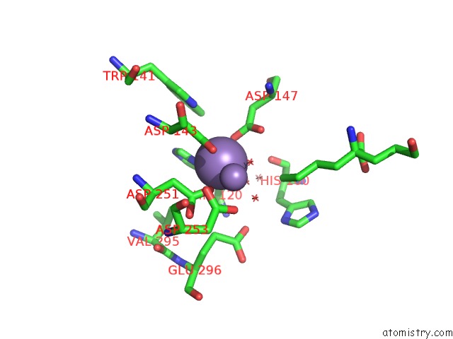
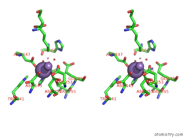
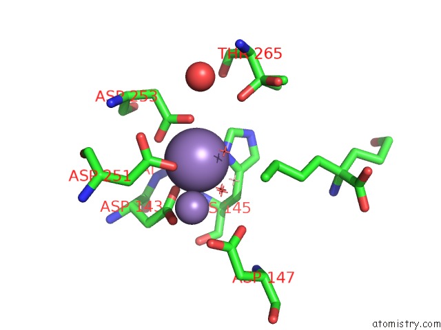
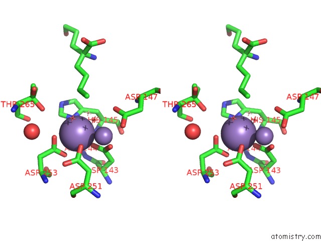
Manganese binding site 1 out of 6 in 4ie2
Go back to
Manganese binding site 1 out
of 6 in the Crystal Structure of Human Arginase-2 Complexed with Inhibitor 1H
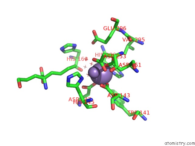
Mono view
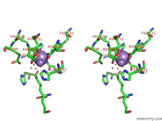
Stereo pair view
Mono view
Stereo pair view
A full contact list of Manganese with other atoms in the Mn binding
site number 1 of Crystal Structure of Human Arginase-2 Complexed with Inhibitor 1H within 5.0Å range:
|
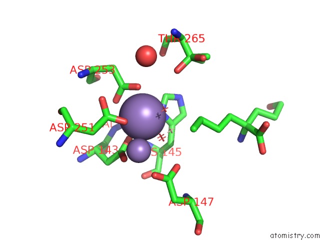
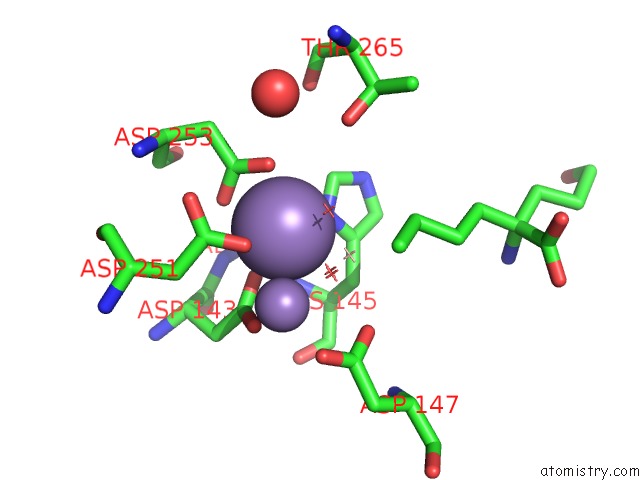
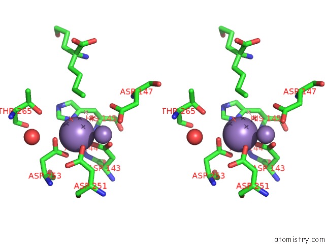
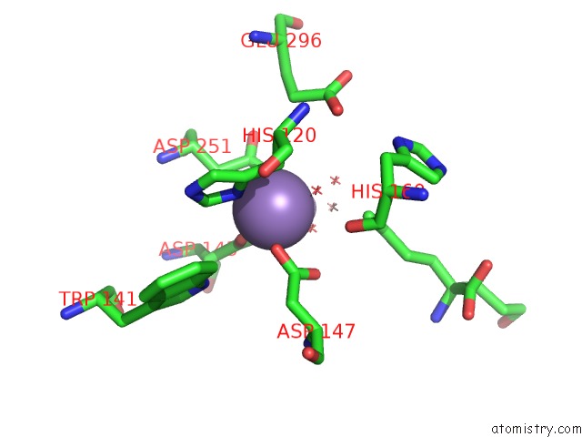
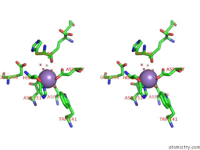
Manganese binding site 2 out of 6 in 4ie2
Go back to
Manganese binding site 2 out
of 6 in the Crystal Structure of Human Arginase-2 Complexed with Inhibitor 1H
Mono view
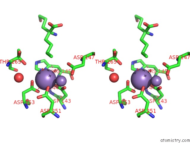
Stereo pair view
Mono view
Stereo pair view
A full contact list of Manganese with other atoms in the Mn binding
site number 2 of Crystal Structure of Human Arginase-2 Complexed with Inhibitor 1H within 5.0Å range:
|
Manganese binding site 3 out of 6 in 4ie2
Go back to
Manganese binding site 3 out
of 6 in the Crystal Structure of Human Arginase-2 Complexed with Inhibitor 1H
Mono view
Stereo pair view
Mono view
Stereo pair view
A full contact list of Manganese with other atoms in the Mn binding
site number 3 of Crystal Structure of Human Arginase-2 Complexed with Inhibitor 1H within 5.0Å range:
|
Manganese binding site 4 out of 6 in 4ie2
Go back to
Manganese binding site 4 out
of 6 in the Crystal Structure of Human Arginase-2 Complexed with Inhibitor 1H
Mono view
Stereo pair view
Mono view
Stereo pair view
A full contact list of Manganese with other atoms in the Mn binding
site number 4 of Crystal Structure of Human Arginase-2 Complexed with Inhibitor 1H within 5.0Å range:
|
Manganese binding site 5 out of 6 in 4ie2
Go back to
Manganese binding site 5 out
of 6 in the Crystal Structure of Human Arginase-2 Complexed with Inhibitor 1H
Mono view
Stereo pair view
Mono view
Stereo pair view
A full contact list of Manganese with other atoms in the Mn binding
site number 5 of Crystal Structure of Human Arginase-2 Complexed with Inhibitor 1H within 5.0Å range:
|
Manganese binding site 6 out of 6 in 4ie2
Go back to
Manganese binding site 6 out
of 6 in the Crystal Structure of Human Arginase-2 Complexed with Inhibitor 1H
Mono view
Stereo pair view
Mono view
Stereo pair view
A full contact list of Manganese with other atoms in the Mn binding
site number 6 of Crystal Structure of Human Arginase-2 Complexed with Inhibitor 1H within 5.0Å range:
|
Reference:
A.Golebiowski,
R.Paul Beckett,
M.Van Zandt,
M.K.Ji,
D.Whitehouse,
T.R.Ryder,
E.Jagdmann,
M.Andreoli,
A.Mazur,
M.Padmanilayam,
A.Cousido-Siah,
A.Mitschler,
F.X.Ruiz,
A.Podjarny,
H.Schroeter.
2-Substituted-2-Amino-6-Boronohexanoic Acids As Arginase Inhibitors. Bioorg.Med.Chem.Lett. V. 23 2027 2013.
ISSN: ISSN 0960-894X
PubMed: 23453840
DOI: 10.1016/J.BMCL.2013.02.024
Page generated: Sat Oct 5 19:47:32 2024
ISSN: ISSN 0960-894X
PubMed: 23453840
DOI: 10.1016/J.BMCL.2013.02.024
Last articles
Zn in 9MJ5Zn in 9HNW
Zn in 9G0L
Zn in 9FNE
Zn in 9DZN
Zn in 9E0I
Zn in 9D32
Zn in 9DAK
Zn in 8ZXC
Zn in 8ZUF