Manganese in PDB 4i06: Crystal Structure of Human Arginase-2 Complexed with Inhibitor 14
Enzymatic activity of Crystal Structure of Human Arginase-2 Complexed with Inhibitor 14
All present enzymatic activity of Crystal Structure of Human Arginase-2 Complexed with Inhibitor 14:
3.5.3.1;
Protein crystallography data
The structure of Crystal Structure of Human Arginase-2 Complexed with Inhibitor 14, PDB code: 4i06
was solved by
A.Cousido-Siah,
A.Mitschler,
F.X.Ruiz,
D.L.Whitehouse,
A.Golebiowski,
M.Ji,
M.Zhang,
P.Beckett,
R.Sheeler,
M.Andreoli,
B.Conway,
K.Mahboubi,
H.Schroeter,
M.C.Van Zandt,
A.Podjarny,
with X-Ray Crystallography technique. A brief refinement statistics is given in the table below:
Resolution Low / High (Å)
|
40.81 /
1.80
|
Space group
|
P 42 21 2
|
Cell size a, b, c (Å), α, β, γ (°)
|
127.805,
127.805,
159.094,
90.00,
90.00,
90.00
|
R / Rfree (%)
|
16.4 /
19.5
|
Manganese Binding Sites:
The binding sites of Manganese atom in the Crystal Structure of Human Arginase-2 Complexed with Inhibitor 14
(pdb code 4i06). This binding sites where shown within
5.0 Angstroms radius around Manganese atom.
In total 6 binding sites of Manganese where determined in the
Crystal Structure of Human Arginase-2 Complexed with Inhibitor 14, PDB code: 4i06:
Jump to Manganese binding site number:
1;
2;
3;
4;
5;
6;
Manganese binding site 1 out
of 6 in 4i06
Go back to
Manganese Binding Sites List in 4i06
Manganese binding site 1 out
of 6 in the Crystal Structure of Human Arginase-2 Complexed with Inhibitor 14
Mono view
Stereo pair view
|
A full contact list of Manganese with other atoms in the Mn binding
site number 1 of Crystal Structure of Human Arginase-2 Complexed with Inhibitor 14 within 5.0Å range:
probe
|
atom
|
residue
|
distance (Å)
|
B
|
Occ
|
A:Mn401
b:11.5
occ:1.00
|
OD2
|
A:ASP147
|
2.1
|
10.1
|
1.0
|
OD2
|
A:ASP143
|
2.1
|
11.3
|
1.0
|
ND1
|
A:HIS120
|
2.2
|
11.9
|
1.0
|
O21
|
A:X8A406
|
2.3
|
10.9
|
1.0
|
O22
|
A:X8A406
|
2.3
|
12.4
|
1.0
|
OD2
|
A:ASP251
|
2.3
|
12.2
|
1.0
|
B19
|
A:X8A406
|
2.8
|
11.7
|
1.0
|
CG
|
A:ASP147
|
3.1
|
11.8
|
1.0
|
CE1
|
A:HIS120
|
3.1
|
12.5
|
1.0
|
CG
|
A:ASP143
|
3.1
|
14.1
|
1.0
|
CG
|
A:HIS120
|
3.2
|
11.8
|
1.0
|
CG
|
A:ASP251
|
3.3
|
11.6
|
1.0
|
MN
|
A:MN402
|
3.4
|
11.7
|
1.0
|
OD1
|
A:ASP147
|
3.4
|
10.0
|
1.0
|
OD1
|
A:ASP143
|
3.4
|
11.0
|
1.0
|
CB
|
A:HIS120
|
3.5
|
9.8
|
1.0
|
O20
|
A:X8A406
|
3.6
|
12.1
|
1.0
|
CB
|
A:ASP251
|
3.6
|
9.5
|
1.0
|
C5
|
A:X8A406
|
4.2
|
12.2
|
1.0
|
NE2
|
A:HIS120
|
4.2
|
10.9
|
1.0
|
NE1
|
A:TRP141
|
4.2
|
12.1
|
1.0
|
CD2
|
A:HIS120
|
4.3
|
9.6
|
1.0
|
OD1
|
A:ASP251
|
4.4
|
9.7
|
1.0
|
CB
|
A:ASP147
|
4.4
|
11.6
|
1.0
|
O
|
A:HIS160
|
4.5
|
10.8
|
1.0
|
CB
|
A:ASP143
|
4.5
|
8.2
|
1.0
|
CZ2
|
A:TRP141
|
4.5
|
12.3
|
1.0
|
CE2
|
A:TRP141
|
4.7
|
13.0
|
1.0
|
CG
|
A:GLU296
|
4.8
|
11.1
|
1.0
|
CA
|
A:ASP251
|
4.9
|
10.6
|
1.0
|
CG2
|
A:VAL295
|
5.0
|
9.5
|
1.0
|
CA
|
A:HIS120
|
5.0
|
9.2
|
1.0
|
|
Manganese binding site 2 out
of 6 in 4i06
Go back to
Manganese Binding Sites List in 4i06
Manganese binding site 2 out
of 6 in the Crystal Structure of Human Arginase-2 Complexed with Inhibitor 14
Mono view
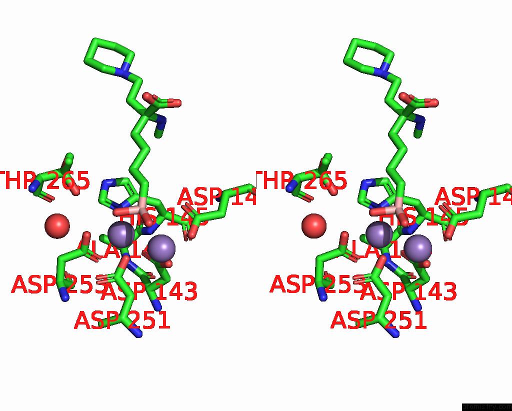
Stereo pair view
|
A full contact list of Manganese with other atoms in the Mn binding
site number 2 of Crystal Structure of Human Arginase-2 Complexed with Inhibitor 14 within 5.0Å range:
probe
|
atom
|
residue
|
distance (Å)
|
B
|
Occ
|
A:Mn402
b:11.7
occ:1.00
|
O22
|
A:X8A406
|
2.1
|
12.4
|
1.0
|
OD1
|
A:ASP143
|
2.2
|
11.0
|
1.0
|
OD2
|
A:ASP253
|
2.2
|
11.9
|
1.0
|
ND1
|
A:HIS145
|
2.2
|
12.5
|
1.0
|
OD2
|
A:ASP251
|
2.3
|
12.2
|
1.0
|
OD1
|
A:ASP253
|
2.4
|
10.8
|
1.0
|
CG
|
A:ASP253
|
2.7
|
14.3
|
1.0
|
O20
|
A:X8A406
|
2.8
|
12.1
|
1.0
|
CE1
|
A:HIS145
|
3.1
|
11.6
|
1.0
|
B19
|
A:X8A406
|
3.1
|
11.7
|
1.0
|
CG
|
A:ASP251
|
3.1
|
11.6
|
1.0
|
CG
|
A:ASP143
|
3.2
|
14.1
|
1.0
|
CG
|
A:HIS145
|
3.3
|
10.2
|
1.0
|
MN
|
A:MN401
|
3.4
|
11.5
|
1.0
|
OD2
|
A:ASP143
|
3.5
|
11.3
|
1.0
|
OD1
|
A:ASP251
|
3.7
|
9.7
|
1.0
|
CB
|
A:HIS145
|
3.7
|
9.4
|
1.0
|
O21
|
A:X8A406
|
4.0
|
10.9
|
1.0
|
OG1
|
A:THR265
|
4.0
|
12.6
|
0.3
|
N
|
A:HIS145
|
4.0
|
9.6
|
1.0
|
CB
|
A:ASP251
|
4.1
|
9.5
|
1.0
|
CB
|
A:ASP253
|
4.2
|
11.0
|
1.0
|
N
|
A:ALA144
|
4.2
|
9.9
|
1.0
|
NE2
|
A:HIS145
|
4.2
|
11.0
|
1.0
|
C5
|
A:X8A406
|
4.3
|
12.2
|
1.0
|
CD2
|
A:HIS145
|
4.4
|
13.7
|
1.0
|
C4
|
A:X8A406
|
4.4
|
12.5
|
1.0
|
OD1
|
A:ASP147
|
4.5
|
10.0
|
1.0
|
CA
|
A:HIS145
|
4.5
|
10.2
|
1.0
|
CB
|
A:ASP143
|
4.6
|
8.2
|
1.0
|
CB
|
A:ALA144
|
4.6
|
10.9
|
1.0
|
O
|
A:HOH513
|
4.6
|
13.2
|
1.0
|
CA
|
A:ALA144
|
4.8
|
11.0
|
1.0
|
OD2
|
A:ASP147
|
4.8
|
10.1
|
1.0
|
C
|
A:ALA144
|
4.8
|
12.4
|
1.0
|
CA
|
A:ASP143
|
4.9
|
9.7
|
1.0
|
C
|
A:ASP143
|
5.0
|
10.6
|
1.0
|
|
Manganese binding site 3 out
of 6 in 4i06
Go back to
Manganese Binding Sites List in 4i06
Manganese binding site 3 out
of 6 in the Crystal Structure of Human Arginase-2 Complexed with Inhibitor 14
Mono view
Stereo pair view
|
A full contact list of Manganese with other atoms in the Mn binding
site number 3 of Crystal Structure of Human Arginase-2 Complexed with Inhibitor 14 within 5.0Å range:
probe
|
atom
|
residue
|
distance (Å)
|
B
|
Occ
|
B:Mn401
b:11.7
occ:1.00
|
OD2
|
B:ASP147
|
2.1
|
12.0
|
1.0
|
OD2
|
B:ASP143
|
2.1
|
11.1
|
1.0
|
OD2
|
B:ASP251
|
2.2
|
10.1
|
1.0
|
O21
|
B:X8A406
|
2.2
|
13.5
|
1.0
|
ND1
|
B:HIS120
|
2.2
|
9.2
|
1.0
|
O22
|
B:X8A406
|
2.4
|
14.7
|
1.0
|
B19
|
B:X8A406
|
2.7
|
13.5
|
1.0
|
CG
|
B:ASP147
|
3.1
|
13.8
|
1.0
|
CG
|
B:ASP143
|
3.1
|
14.7
|
1.0
|
CE1
|
B:HIS120
|
3.2
|
12.2
|
1.0
|
CG
|
B:HIS120
|
3.2
|
12.0
|
1.0
|
CG
|
B:ASP251
|
3.2
|
11.2
|
1.0
|
MN
|
B:MN402
|
3.3
|
12.0
|
1.0
|
OD1
|
B:ASP143
|
3.4
|
11.9
|
1.0
|
OD1
|
B:ASP147
|
3.4
|
12.4
|
1.0
|
CB
|
B:HIS120
|
3.5
|
12.4
|
1.0
|
O20
|
B:X8A406
|
3.6
|
13.4
|
1.0
|
CB
|
B:ASP251
|
3.6
|
11.1
|
1.0
|
C5
|
B:X8A406
|
4.1
|
12.6
|
1.0
|
NE1
|
B:TRP141
|
4.3
|
9.8
|
1.0
|
NE2
|
B:HIS120
|
4.3
|
10.6
|
1.0
|
CD2
|
B:HIS120
|
4.3
|
9.1
|
1.0
|
OD1
|
B:ASP251
|
4.3
|
11.6
|
1.0
|
O
|
B:HIS160
|
4.4
|
10.6
|
1.0
|
CB
|
B:ASP147
|
4.5
|
10.4
|
1.0
|
CB
|
B:ASP143
|
4.5
|
9.0
|
1.0
|
CZ2
|
B:TRP141
|
4.6
|
11.1
|
1.0
|
CG
|
B:GLU296
|
4.8
|
10.0
|
1.0
|
CE2
|
B:TRP141
|
4.8
|
10.9
|
1.0
|
CA
|
B:ASP251
|
4.9
|
9.5
|
1.0
|
CG2
|
B:VAL295
|
4.9
|
10.0
|
1.0
|
CA
|
B:HIS120
|
5.0
|
9.0
|
1.0
|
OD2
|
B:ASP253
|
5.0
|
9.8
|
1.0
|
|
Manganese binding site 4 out
of 6 in 4i06
Go back to
Manganese Binding Sites List in 4i06
Manganese binding site 4 out
of 6 in the Crystal Structure of Human Arginase-2 Complexed with Inhibitor 14
Mono view
Stereo pair view
|
A full contact list of Manganese with other atoms in the Mn binding
site number 4 of Crystal Structure of Human Arginase-2 Complexed with Inhibitor 14 within 5.0Å range:
probe
|
atom
|
residue
|
distance (Å)
|
B
|
Occ
|
B:Mn402
b:12.0
occ:1.00
|
O22
|
B:X8A406
|
2.0
|
14.7
|
1.0
|
OD1
|
B:ASP143
|
2.1
|
11.9
|
1.0
|
OD2
|
B:ASP253
|
2.2
|
9.8
|
1.0
|
ND1
|
B:HIS145
|
2.3
|
10.3
|
1.0
|
OD2
|
B:ASP251
|
2.4
|
10.1
|
1.0
|
OD1
|
B:ASP253
|
2.4
|
11.6
|
1.0
|
CG
|
B:ASP253
|
2.6
|
10.8
|
1.0
|
O20
|
B:X8A406
|
2.8
|
13.4
|
1.0
|
B19
|
B:X8A406
|
3.1
|
13.5
|
1.0
|
CE1
|
B:HIS145
|
3.1
|
12.4
|
1.0
|
CG
|
B:ASP251
|
3.1
|
11.2
|
1.0
|
CG
|
B:ASP143
|
3.2
|
14.7
|
1.0
|
MN
|
B:MN401
|
3.3
|
11.7
|
1.0
|
CG
|
B:HIS145
|
3.4
|
13.3
|
1.0
|
OD2
|
B:ASP143
|
3.5
|
11.1
|
1.0
|
OD1
|
B:ASP251
|
3.6
|
11.6
|
1.0
|
CB
|
B:HIS145
|
3.8
|
12.5
|
1.0
|
O21
|
B:X8A406
|
3.9
|
13.5
|
1.0
|
N
|
B:HIS145
|
4.0
|
11.6
|
1.0
|
CB
|
B:ASP251
|
4.1
|
11.1
|
1.0
|
OG1
|
B:THR265
|
4.1
|
39.1
|
1.0
|
CB
|
B:ASP253
|
4.1
|
8.3
|
1.0
|
N
|
B:ALA144
|
4.2
|
11.8
|
1.0
|
C5
|
B:X8A406
|
4.3
|
12.6
|
1.0
|
NE2
|
B:HIS145
|
4.3
|
10.9
|
1.0
|
C4
|
B:X8A406
|
4.4
|
13.8
|
1.0
|
CD2
|
B:HIS145
|
4.4
|
13.8
|
1.0
|
CA
|
B:HIS145
|
4.5
|
10.1
|
1.0
|
CB
|
B:ASP143
|
4.5
|
9.0
|
1.0
|
OD1
|
B:ASP147
|
4.5
|
12.4
|
1.0
|
CB
|
B:ALA144
|
4.6
|
12.0
|
1.0
|
O
|
B:HOH526
|
4.6
|
14.0
|
1.0
|
CA
|
B:ALA144
|
4.7
|
11.1
|
1.0
|
C
|
B:ALA144
|
4.8
|
14.2
|
1.0
|
OD2
|
B:ASP147
|
4.8
|
12.0
|
1.0
|
CA
|
B:ASP143
|
4.9
|
9.4
|
1.0
|
C
|
B:ASP143
|
5.0
|
9.7
|
1.0
|
|
Manganese binding site 5 out
of 6 in 4i06
Go back to
Manganese Binding Sites List in 4i06
Manganese binding site 5 out
of 6 in the Crystal Structure of Human Arginase-2 Complexed with Inhibitor 14
Mono view
Stereo pair view
|
A full contact list of Manganese with other atoms in the Mn binding
site number 5 of Crystal Structure of Human Arginase-2 Complexed with Inhibitor 14 within 5.0Å range:
probe
|
atom
|
residue
|
distance (Å)
|
B
|
Occ
|
C:Mn401
b:11.7
occ:1.00
|
O22
|
C:X8A406
|
2.1
|
13.0
|
1.0
|
OD1
|
C:ASP143
|
2.2
|
10.2
|
1.0
|
ND1
|
C:HIS145
|
2.2
|
11.7
|
1.0
|
OD2
|
C:ASP253
|
2.2
|
10.5
|
1.0
|
OD2
|
C:ASP251
|
2.2
|
11.2
|
1.0
|
OD1
|
C:ASP253
|
2.4
|
11.8
|
1.0
|
CG
|
C:ASP253
|
2.7
|
13.7
|
1.0
|
O20
|
C:X8A406
|
2.7
|
14.2
|
1.0
|
CE1
|
C:HIS145
|
3.1
|
13.6
|
1.0
|
B19
|
C:X8A406
|
3.1
|
16.1
|
1.0
|
CG
|
C:ASP251
|
3.1
|
13.3
|
1.0
|
CG
|
C:ASP143
|
3.2
|
9.8
|
1.0
|
CG
|
C:HIS145
|
3.3
|
9.7
|
1.0
|
MN
|
C:MN402
|
3.4
|
11.8
|
1.0
|
OD2
|
C:ASP143
|
3.5
|
12.1
|
1.0
|
CB
|
C:HIS145
|
3.7
|
12.6
|
1.0
|
OD1
|
C:ASP251
|
3.7
|
11.0
|
1.0
|
O21
|
C:X8A406
|
4.0
|
12.7
|
1.0
|
OG1
|
C:THR265
|
4.0
|
13.5
|
0.2
|
N
|
C:HIS145
|
4.1
|
11.7
|
1.0
|
CB
|
C:ASP251
|
4.1
|
12.5
|
1.0
|
CB
|
C:ASP253
|
4.2
|
9.8
|
1.0
|
NE2
|
C:HIS145
|
4.2
|
10.9
|
1.0
|
N
|
C:ALA144
|
4.3
|
11.2
|
1.0
|
C5
|
C:X8A406
|
4.3
|
10.6
|
1.0
|
C4
|
C:X8A406
|
4.3
|
13.5
|
1.0
|
CD2
|
C:HIS145
|
4.4
|
11.0
|
1.0
|
OD1
|
C:ASP147
|
4.5
|
12.1
|
1.0
|
CA
|
C:HIS145
|
4.5
|
10.2
|
1.0
|
CB
|
C:ASP143
|
4.6
|
11.9
|
1.0
|
O
|
C:HOH526
|
4.6
|
13.8
|
1.0
|
CB
|
C:ALA144
|
4.7
|
12.9
|
1.0
|
C
|
C:ALA144
|
4.8
|
9.8
|
1.0
|
CA
|
C:ALA144
|
4.8
|
9.4
|
1.0
|
OD2
|
C:ASP147
|
4.8
|
12.9
|
1.0
|
CA
|
C:ASP143
|
4.9
|
10.8
|
1.0
|
O
|
C:HOH549
|
5.0
|
18.1
|
1.0
|
|
Manganese binding site 6 out
of 6 in 4i06
Go back to
Manganese Binding Sites List in 4i06
Manganese binding site 6 out
of 6 in the Crystal Structure of Human Arginase-2 Complexed with Inhibitor 14
Mono view
Stereo pair view
|
A full contact list of Manganese with other atoms in the Mn binding
site number 6 of Crystal Structure of Human Arginase-2 Complexed with Inhibitor 14 within 5.0Å range:
probe
|
atom
|
residue
|
distance (Å)
|
B
|
Occ
|
C:Mn402
b:11.8
occ:1.00
|
OD2
|
C:ASP147
|
2.1
|
12.9
|
1.0
|
ND1
|
C:HIS120
|
2.1
|
10.6
|
1.0
|
OD2
|
C:ASP143
|
2.1
|
12.1
|
1.0
|
O22
|
C:X8A406
|
2.2
|
13.0
|
1.0
|
O21
|
C:X8A406
|
2.3
|
12.7
|
1.0
|
OD2
|
C:ASP251
|
2.3
|
11.2
|
1.0
|
B19
|
C:X8A406
|
2.7
|
16.1
|
1.0
|
CG
|
C:ASP147
|
3.1
|
13.8
|
1.0
|
CE1
|
C:HIS120
|
3.1
|
13.1
|
1.0
|
CG
|
C:HIS120
|
3.1
|
12.2
|
1.0
|
CG
|
C:ASP143
|
3.1
|
9.8
|
1.0
|
CG
|
C:ASP251
|
3.3
|
13.3
|
1.0
|
OD1
|
C:ASP147
|
3.3
|
12.1
|
1.0
|
MN
|
C:MN401
|
3.4
|
11.7
|
1.0
|
CB
|
C:HIS120
|
3.5
|
10.5
|
1.0
|
OD1
|
C:ASP143
|
3.5
|
10.2
|
1.0
|
O20
|
C:X8A406
|
3.5
|
14.2
|
1.0
|
CB
|
C:ASP251
|
3.6
|
12.5
|
1.0
|
C5
|
C:X8A406
|
4.1
|
10.6
|
1.0
|
NE2
|
C:HIS120
|
4.2
|
14.1
|
1.0
|
CD2
|
C:HIS120
|
4.2
|
12.7
|
1.0
|
NE1
|
C:TRP141
|
4.3
|
11.6
|
1.0
|
OD1
|
C:ASP251
|
4.4
|
11.0
|
1.0
|
CB
|
C:ASP147
|
4.5
|
10.7
|
1.0
|
O
|
C:HIS160
|
4.5
|
12.3
|
1.0
|
CB
|
C:ASP143
|
4.5
|
11.9
|
1.0
|
CZ2
|
C:TRP141
|
4.6
|
11.7
|
1.0
|
CE2
|
C:TRP141
|
4.8
|
14.4
|
1.0
|
CG
|
C:GLU296
|
4.8
|
10.4
|
1.0
|
CA
|
C:ASP251
|
4.9
|
12.0
|
1.0
|
CG2
|
C:VAL295
|
4.9
|
12.3
|
1.0
|
CA
|
C:HIS120
|
5.0
|
13.8
|
1.0
|
CB
|
C:HIS145
|
5.0
|
12.6
|
1.0
|
|
Reference:
M.C.Van Zandt,
D.L.Whitehouse,
A.Golebiowski,
M.K.Ji,
M.Zhang,
R.P.Beckett,
G.E.Jagdmann,
T.R.Ryder,
R.Sheeler,
M.Andreoli,
B.Conway,
K.Mahboubi,
G.D'angelo,
A.Mitschler,
A.Cousido-Siah,
F.X.Ruiz,
E.I.Howard,
A.D.Podjarny,
H.Schroeter.
Discovery of (R)-2-Amino-6-Borono-2-(2-(Piperidin-1-Yl)Ethyl)Hexanoic Acid and Congeners As Highly Potent Inhibitors of Human Arginases I and II For Treatment of Myocardial Reperfusion Injury. J.Med.Chem. V. 56 2568 2013.
ISSN: ISSN 0022-2623
PubMed: 23472952
DOI: 10.1021/JM400014C
Page generated: Sat Oct 5 19:45:24 2024
|