Manganese »
PDB 4gwc-4ilk »
4hnt »
Manganese in PDB 4hnt: Crystal Structure of F403A Mutant of S. Aureus Pyruvate Carboxylase
Enzymatic activity of Crystal Structure of F403A Mutant of S. Aureus Pyruvate Carboxylase
All present enzymatic activity of Crystal Structure of F403A Mutant of S. Aureus Pyruvate Carboxylase:
6.4.1.1;
6.4.1.1;
Protein crystallography data
The structure of Crystal Structure of F403A Mutant of S. Aureus Pyruvate Carboxylase, PDB code: 4hnt
was solved by
L.P.C.Yu,
L.Tong,
with X-Ray Crystallography technique. A brief refinement statistics is given in the table below:
| Resolution Low / High (Å) | 30.00 / 2.80 |
| Space group | P 1 21 1 |
| Cell size a, b, c (Å), α, β, γ (°) | 96.230, 256.283, 126.685, 90.00, 109.86, 90.00 |
| R / Rfree (%) | 20.9 / 27.9 |
Manganese Binding Sites:
The binding sites of Manganese atom in the Crystal Structure of F403A Mutant of S. Aureus Pyruvate Carboxylase
(pdb code 4hnt). This binding sites where shown within
5.0 Angstroms radius around Manganese atom.
In total 4 binding sites of Manganese where determined in the Crystal Structure of F403A Mutant of S. Aureus Pyruvate Carboxylase, PDB code: 4hnt:
Jump to Manganese binding site number: 1; 2; 3; 4;
In total 4 binding sites of Manganese where determined in the Crystal Structure of F403A Mutant of S. Aureus Pyruvate Carboxylase, PDB code: 4hnt:
Jump to Manganese binding site number: 1; 2; 3; 4;
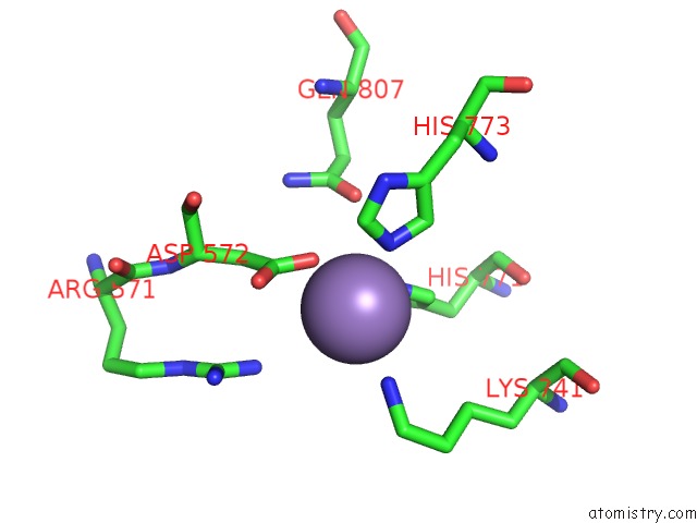
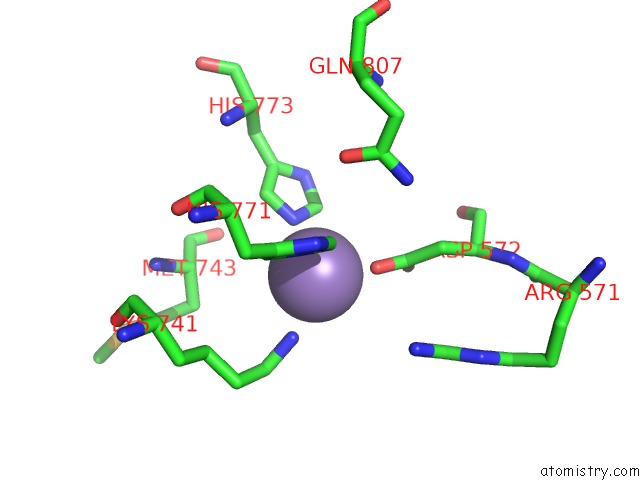
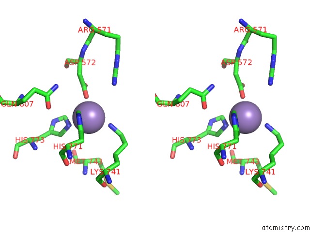
Manganese binding site 1 out of 4 in 4hnt
Go back to
Manganese binding site 1 out
of 4 in the Crystal Structure of F403A Mutant of S. Aureus Pyruvate Carboxylase
Mono view
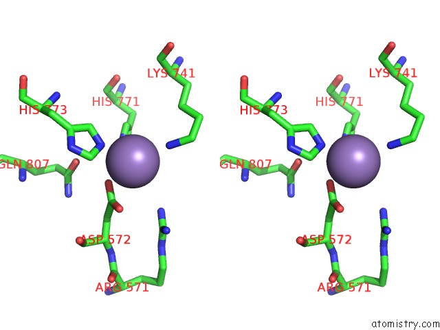
Stereo pair view
Mono view
Stereo pair view
A full contact list of Manganese with other atoms in the Mn binding
site number 1 of Crystal Structure of F403A Mutant of S. Aureus Pyruvate Carboxylase within 5.0Å range:
|
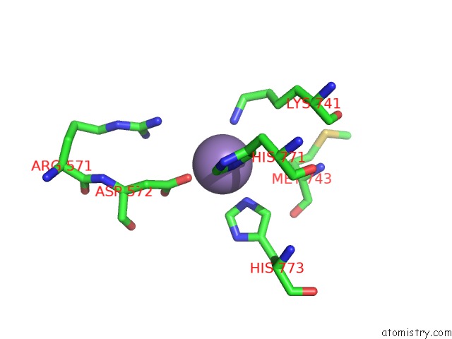
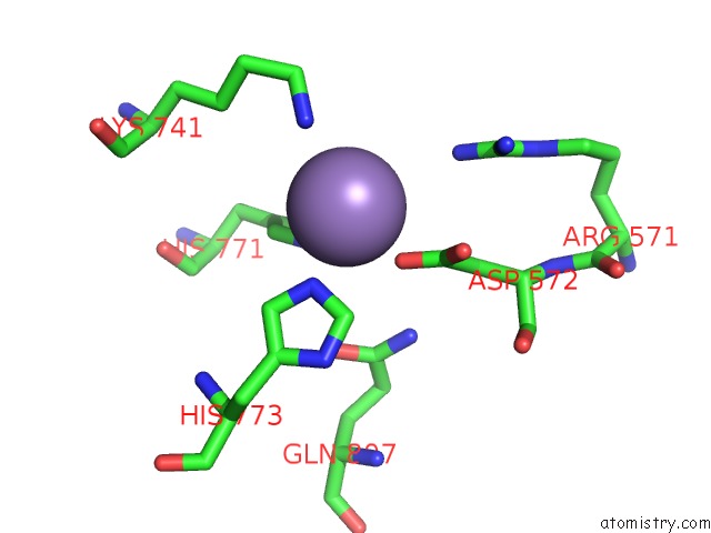
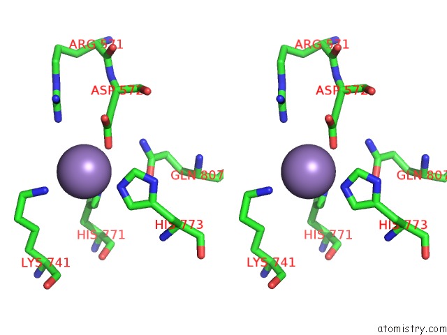
Manganese binding site 2 out of 4 in 4hnt
Go back to
Manganese binding site 2 out
of 4 in the Crystal Structure of F403A Mutant of S. Aureus Pyruvate Carboxylase
Mono view
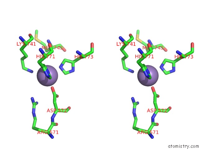
Stereo pair view
Mono view
Stereo pair view
A full contact list of Manganese with other atoms in the Mn binding
site number 2 of Crystal Structure of F403A Mutant of S. Aureus Pyruvate Carboxylase within 5.0Å range:
|
Manganese binding site 3 out of 4 in 4hnt
Go back to
Manganese binding site 3 out
of 4 in the Crystal Structure of F403A Mutant of S. Aureus Pyruvate Carboxylase
Mono view
Stereo pair view
Mono view
Stereo pair view
A full contact list of Manganese with other atoms in the Mn binding
site number 3 of Crystal Structure of F403A Mutant of S. Aureus Pyruvate Carboxylase within 5.0Å range:
|
Manganese binding site 4 out of 4 in 4hnt
Go back to
Manganese binding site 4 out
of 4 in the Crystal Structure of F403A Mutant of S. Aureus Pyruvate Carboxylase
Mono view
Stereo pair view
Mono view
Stereo pair view
A full contact list of Manganese with other atoms in the Mn binding
site number 4 of Crystal Structure of F403A Mutant of S. Aureus Pyruvate Carboxylase within 5.0Å range:
|
Reference:
L.P.Yu,
C.Y.Chou,
P.H.Choi,
L.Tong.
Characterizing the Importance of the Biotin Carboxylase Domain Dimer For Staphylococcus Aureus Pyruvate Carboxylase Catalysis. Biochemistry V. 52 488 2013.
ISSN: ISSN 0006-2960
PubMed: 23286247
DOI: 10.1021/BI301294D
Page generated: Sat Oct 5 19:42:26 2024
ISSN: ISSN 0006-2960
PubMed: 23286247
DOI: 10.1021/BI301294D
Last articles
Zn in 9MJ5Zn in 9HNW
Zn in 9G0L
Zn in 9FNE
Zn in 9DZN
Zn in 9E0I
Zn in 9D32
Zn in 9DAK
Zn in 8ZXC
Zn in 8ZUF