Manganese »
PDB 4gwc-4ilk »
4h55 »
Manganese in PDB 4h55: Crystal Structure of Canavalia Brasiliensis Seed Lectin (Conbr) in Complex with Beta-D-Ribofuranose
Protein crystallography data
The structure of Crystal Structure of Canavalia Brasiliensis Seed Lectin (Conbr) in Complex with Beta-D-Ribofuranose, PDB code: 4h55
was solved by
E.Salviano,
B.A.M.Rocha,
B.S.Cavada,
P.Delatorre,
T.Santi-Gadelha,
C.A.A.Gadelha,
J.C.Silva-Filho,
R.B.Nobrega,
D.L.Farias,
with X-Ray Crystallography technique. A brief refinement statistics is given in the table below:
| Resolution Low / High (Å) | 24.39 / 2.15 |
| Space group | I 2 2 2 |
| Cell size a, b, c (Å), α, β, γ (°) | 67.240, 70.430, 97.570, 90.00, 90.00, 90.00 |
| R / Rfree (%) | 18.3 / 22.7 |
Other elements in 4h55:
The structure of Crystal Structure of Canavalia Brasiliensis Seed Lectin (Conbr) in Complex with Beta-D-Ribofuranose also contains other interesting chemical elements:
| Calcium | (Ca) | 1 atom |
Manganese Binding Sites:
The binding sites of Manganese atom in the Crystal Structure of Canavalia Brasiliensis Seed Lectin (Conbr) in Complex with Beta-D-Ribofuranose
(pdb code 4h55). This binding sites where shown within
5.0 Angstroms radius around Manganese atom.
In total only one binding site of Manganese was determined in the Crystal Structure of Canavalia Brasiliensis Seed Lectin (Conbr) in Complex with Beta-D-Ribofuranose, PDB code: 4h55:
In total only one binding site of Manganese was determined in the Crystal Structure of Canavalia Brasiliensis Seed Lectin (Conbr) in Complex with Beta-D-Ribofuranose, PDB code: 4h55:
Manganese binding site 1 out of 1 in 4h55
Go back to
Manganese binding site 1 out
of 1 in the Crystal Structure of Canavalia Brasiliensis Seed Lectin (Conbr) in Complex with Beta-D-Ribofuranose
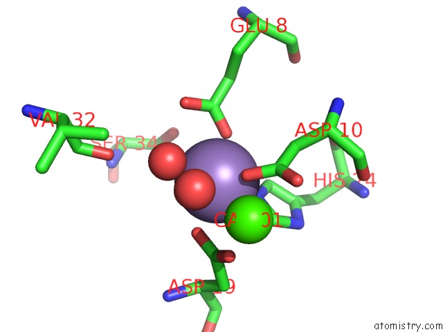
Mono view
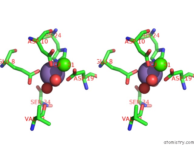
Stereo pair view
Mono view
Stereo pair view
A full contact list of Manganese with other atoms in the Mn binding
site number 1 of Crystal Structure of Canavalia Brasiliensis Seed Lectin (Conbr) in Complex with Beta-D-Ribofuranose within 5.0Å range:
|
Reference:
E.Salviano,
B.A.M.Rocha,
B.S.Cavada,
P.Delatorre,
T.Santi-Gadelha,
C.A.A.Gadelha,
J.C.Silva-Filho,
R.B.Nobrega,
D.L.Farias.
Crystal Structure of Canavalia Brasiliensis Seed Lectin (Conbr) in Complex with Beta-D-Ribofuranose To Be Published.
Page generated: Sat Oct 5 19:40:14 2024
Last articles
Ca in 5TI8Ca in 5THG
Ca in 5TGX
Ca in 5TFK
Ca in 5TG3
Ca in 5TGQ
Ca in 5TGF
Ca in 5TFM
Ca in 5TEA
Ca in 5TFL