Manganese »
PDB 4fo7-4gv9 »
4gnq »
Manganese in PDB 4gnq: Structure of Rat Cytosolic Pepck LD_3G in Complex with Oxalate and Gtp
Enzymatic activity of Structure of Rat Cytosolic Pepck LD_3G in Complex with Oxalate and Gtp
All present enzymatic activity of Structure of Rat Cytosolic Pepck LD_3G in Complex with Oxalate and Gtp:
4.1.1.32;
4.1.1.32;
Protein crystallography data
The structure of Structure of Rat Cytosolic Pepck LD_3G in Complex with Oxalate and Gtp, PDB code: 4gnq
was solved by
T.A.Johnson,
T.Holyoak,
with X-Ray Crystallography technique. A brief refinement statistics is given in the table below:
| Resolution Low / High (Å) | 27.36 / 1.40 |
| Space group | P 21 21 21 |
| Cell size a, b, c (Å), α, β, γ (°) | 60.487, 84.329, 118.971, 90.00, 90.00, 90.00 |
| R / Rfree (%) | 17.4 / 19.7 |
Other elements in 4gnq:
The structure of Structure of Rat Cytosolic Pepck LD_3G in Complex with Oxalate and Gtp also contains other interesting chemical elements:
| Sodium | (Na) | 1 atom |
Manganese Binding Sites:
The binding sites of Manganese atom in the Structure of Rat Cytosolic Pepck LD_3G in Complex with Oxalate and Gtp
(pdb code 4gnq). This binding sites where shown within
5.0 Angstroms radius around Manganese atom.
In total 2 binding sites of Manganese where determined in the Structure of Rat Cytosolic Pepck LD_3G in Complex with Oxalate and Gtp, PDB code: 4gnq:
Jump to Manganese binding site number: 1; 2;
In total 2 binding sites of Manganese where determined in the Structure of Rat Cytosolic Pepck LD_3G in Complex with Oxalate and Gtp, PDB code: 4gnq:
Jump to Manganese binding site number: 1; 2;
Manganese binding site 1 out of 2 in 4gnq
Go back to
Manganese binding site 1 out
of 2 in the Structure of Rat Cytosolic Pepck LD_3G in Complex with Oxalate and Gtp
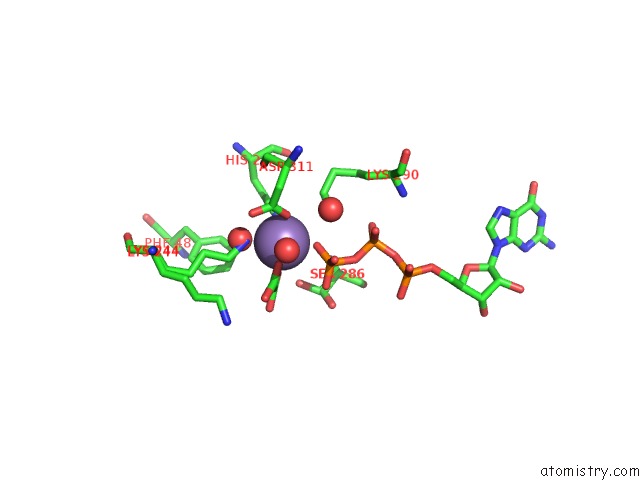
Mono view
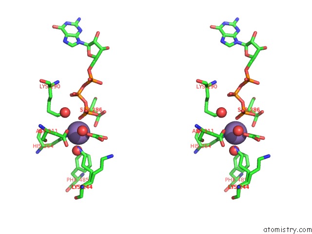
Stereo pair view
Mono view
Stereo pair view
A full contact list of Manganese with other atoms in the Mn binding
site number 1 of Structure of Rat Cytosolic Pepck LD_3G in Complex with Oxalate and Gtp within 5.0Å range:
|
Manganese binding site 2 out of 2 in 4gnq
Go back to
Manganese binding site 2 out
of 2 in the Structure of Rat Cytosolic Pepck LD_3G in Complex with Oxalate and Gtp
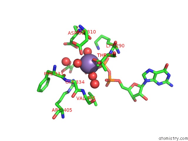
Mono view
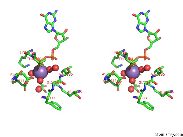
Stereo pair view
Mono view
Stereo pair view
A full contact list of Manganese with other atoms in the Mn binding
site number 2 of Structure of Rat Cytosolic Pepck LD_3G in Complex with Oxalate and Gtp within 5.0Å range:
|
Reference:
T.A.Johnson,
T.Holyoak.
The {Omega}-Loop Lid Domain of Phosphoenolpyruvate Carboxykinase Is Essential For Catalytic Function. Biochemistry V. 51 9547 2012.
ISSN: ISSN 0006-2960
PubMed: 23127136
DOI: 10.1021/BI301278T
Page generated: Sat Oct 5 19:36:20 2024
ISSN: ISSN 0006-2960
PubMed: 23127136
DOI: 10.1021/BI301278T
Last articles
Zn in 9J0NZn in 9J0O
Zn in 9J0P
Zn in 9FJX
Zn in 9EKB
Zn in 9C0F
Zn in 9CAH
Zn in 9CH0
Zn in 9CH3
Zn in 9CH1