Manganese »
PDB 4fo7-4gv9 »
4gjj »
Manganese in PDB 4gjj: Crystal Structure of Pseudomonas Stutzeri L-Rhamnose Isomerase Mutant H101N in Complex with D-Allopyranose
Enzymatic activity of Crystal Structure of Pseudomonas Stutzeri L-Rhamnose Isomerase Mutant H101N in Complex with D-Allopyranose
All present enzymatic activity of Crystal Structure of Pseudomonas Stutzeri L-Rhamnose Isomerase Mutant H101N in Complex with D-Allopyranose:
5.3.1.14;
5.3.1.14;
Protein crystallography data
The structure of Crystal Structure of Pseudomonas Stutzeri L-Rhamnose Isomerase Mutant H101N in Complex with D-Allopyranose, PDB code: 4gjj
was solved by
H.Yoshida,
S.Kamitori,
with X-Ray Crystallography technique. A brief refinement statistics is given in the table below:
| Resolution Low / High (Å) | 37.60 / 2.38 |
| Space group | P 1 21 1 |
| Cell size a, b, c (Å), α, β, γ (°) | 74.650, 104.381, 113.972, 90.00, 107.91, 90.00 |
| R / Rfree (%) | 19.6 / 24.9 |
Manganese Binding Sites:
The binding sites of Manganese atom in the Crystal Structure of Pseudomonas Stutzeri L-Rhamnose Isomerase Mutant H101N in Complex with D-Allopyranose
(pdb code 4gjj). This binding sites where shown within
5.0 Angstroms radius around Manganese atom.
In total 10 binding sites of Manganese where determined in the Crystal Structure of Pseudomonas Stutzeri L-Rhamnose Isomerase Mutant H101N in Complex with D-Allopyranose, PDB code: 4gjj:
Jump to Manganese binding site number: 1; 2; 3; 4; 5; 6; 7; 8; 9; 10;
In total 10 binding sites of Manganese where determined in the Crystal Structure of Pseudomonas Stutzeri L-Rhamnose Isomerase Mutant H101N in Complex with D-Allopyranose, PDB code: 4gjj:
Jump to Manganese binding site number: 1; 2; 3; 4; 5; 6; 7; 8; 9; 10;
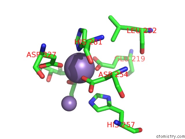
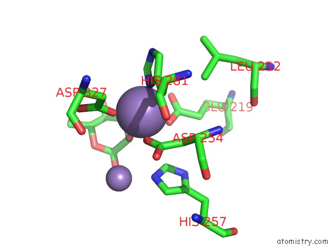
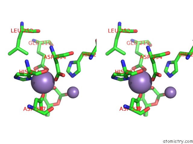
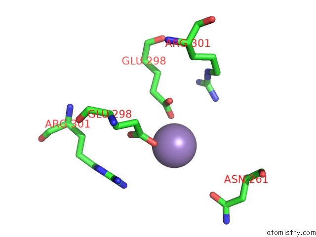
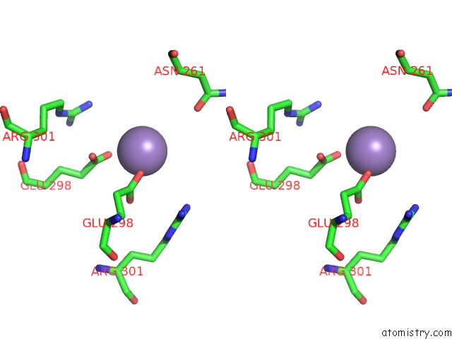
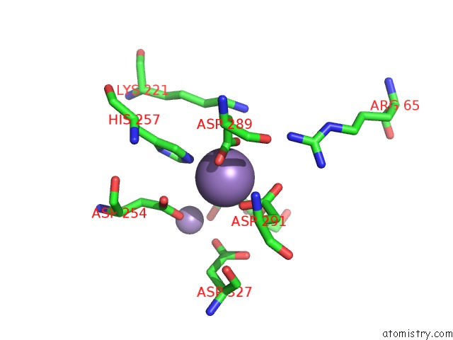
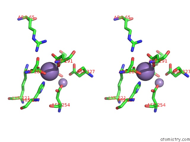
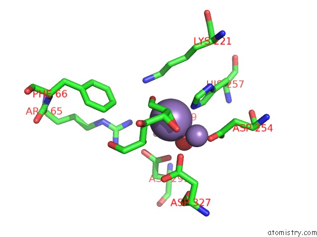
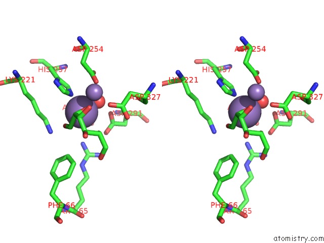
Manganese binding site 1 out of 10 in 4gjj
Go back to
Manganese binding site 1 out
of 10 in the Crystal Structure of Pseudomonas Stutzeri L-Rhamnose Isomerase Mutant H101N in Complex with D-Allopyranose
Mono view
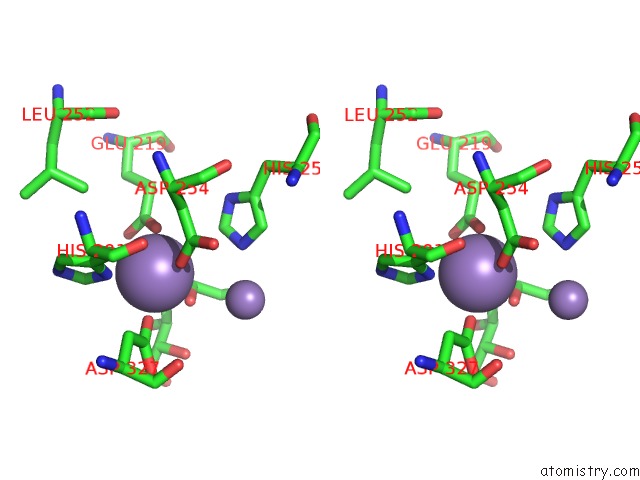
Stereo pair view
Mono view
Stereo pair view
A full contact list of Manganese with other atoms in the Mn binding
site number 1 of Crystal Structure of Pseudomonas Stutzeri L-Rhamnose Isomerase Mutant H101N in Complex with D-Allopyranose within 5.0Å range:
|
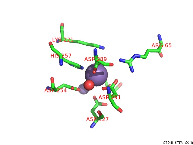
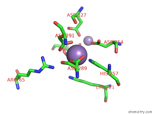
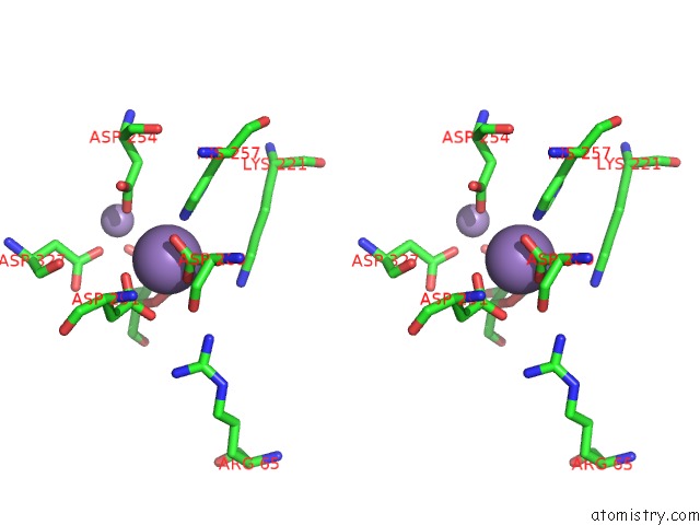
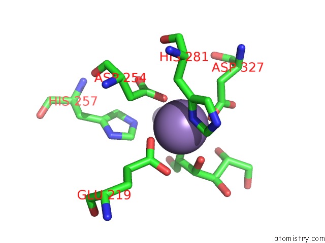
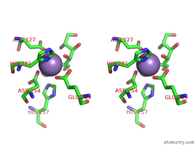
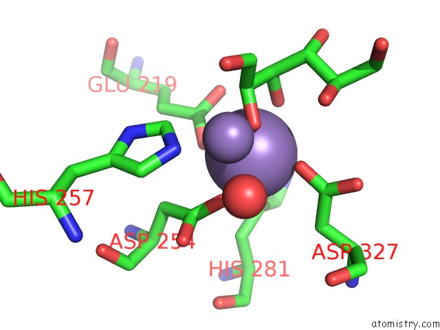
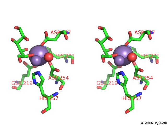
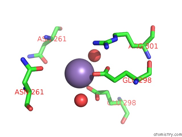
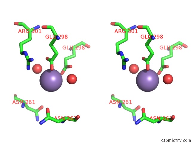
Manganese binding site 2 out of 10 in 4gjj
Go back to
Manganese binding site 2 out
of 10 in the Crystal Structure of Pseudomonas Stutzeri L-Rhamnose Isomerase Mutant H101N in Complex with D-Allopyranose
Mono view
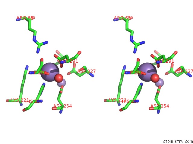
Stereo pair view
Mono view
Stereo pair view
A full contact list of Manganese with other atoms in the Mn binding
site number 2 of Crystal Structure of Pseudomonas Stutzeri L-Rhamnose Isomerase Mutant H101N in Complex with D-Allopyranose within 5.0Å range:
|
Manganese binding site 3 out of 10 in 4gjj
Go back to
Manganese binding site 3 out
of 10 in the Crystal Structure of Pseudomonas Stutzeri L-Rhamnose Isomerase Mutant H101N in Complex with D-Allopyranose
Mono view
Stereo pair view
Mono view
Stereo pair view
A full contact list of Manganese with other atoms in the Mn binding
site number 3 of Crystal Structure of Pseudomonas Stutzeri L-Rhamnose Isomerase Mutant H101N in Complex with D-Allopyranose within 5.0Å range:
|
Manganese binding site 4 out of 10 in 4gjj
Go back to
Manganese binding site 4 out
of 10 in the Crystal Structure of Pseudomonas Stutzeri L-Rhamnose Isomerase Mutant H101N in Complex with D-Allopyranose
Mono view
Stereo pair view
Mono view
Stereo pair view
A full contact list of Manganese with other atoms in the Mn binding
site number 4 of Crystal Structure of Pseudomonas Stutzeri L-Rhamnose Isomerase Mutant H101N in Complex with D-Allopyranose within 5.0Å range:
|
Manganese binding site 5 out of 10 in 4gjj
Go back to
Manganese binding site 5 out
of 10 in the Crystal Structure of Pseudomonas Stutzeri L-Rhamnose Isomerase Mutant H101N in Complex with D-Allopyranose
Mono view
Stereo pair view
Mono view
Stereo pair view
A full contact list of Manganese with other atoms in the Mn binding
site number 5 of Crystal Structure of Pseudomonas Stutzeri L-Rhamnose Isomerase Mutant H101N in Complex with D-Allopyranose within 5.0Å range:
|
Manganese binding site 6 out of 10 in 4gjj
Go back to
Manganese binding site 6 out
of 10 in the Crystal Structure of Pseudomonas Stutzeri L-Rhamnose Isomerase Mutant H101N in Complex with D-Allopyranose
Mono view
Stereo pair view
Mono view
Stereo pair view
A full contact list of Manganese with other atoms in the Mn binding
site number 6 of Crystal Structure of Pseudomonas Stutzeri L-Rhamnose Isomerase Mutant H101N in Complex with D-Allopyranose within 5.0Å range:
|
Manganese binding site 7 out of 10 in 4gjj
Go back to
Manganese binding site 7 out
of 10 in the Crystal Structure of Pseudomonas Stutzeri L-Rhamnose Isomerase Mutant H101N in Complex with D-Allopyranose
Mono view
Stereo pair view
Mono view
Stereo pair view
A full contact list of Manganese with other atoms in the Mn binding
site number 7 of Crystal Structure of Pseudomonas Stutzeri L-Rhamnose Isomerase Mutant H101N in Complex with D-Allopyranose within 5.0Å range:
|
Manganese binding site 8 out of 10 in 4gjj
Go back to
Manganese binding site 8 out
of 10 in the Crystal Structure of Pseudomonas Stutzeri L-Rhamnose Isomerase Mutant H101N in Complex with D-Allopyranose
Mono view
Stereo pair view
Mono view
Stereo pair view
A full contact list of Manganese with other atoms in the Mn binding
site number 8 of Crystal Structure of Pseudomonas Stutzeri L-Rhamnose Isomerase Mutant H101N in Complex with D-Allopyranose within 5.0Å range:
|
Manganese binding site 9 out of 10 in 4gjj
Go back to
Manganese binding site 9 out
of 10 in the Crystal Structure of Pseudomonas Stutzeri L-Rhamnose Isomerase Mutant H101N in Complex with D-Allopyranose
Mono view
Stereo pair view
Mono view
Stereo pair view
A full contact list of Manganese with other atoms in the Mn binding
site number 9 of Crystal Structure of Pseudomonas Stutzeri L-Rhamnose Isomerase Mutant H101N in Complex with D-Allopyranose within 5.0Å range:
|
Manganese binding site 10 out of 10 in 4gjj
Go back to
Manganese binding site 10 out
of 10 in the Crystal Structure of Pseudomonas Stutzeri L-Rhamnose Isomerase Mutant H101N in Complex with D-Allopyranose
Mono view
Stereo pair view
Mono view
Stereo pair view
A full contact list of Manganese with other atoms in the Mn binding
site number 10 of Crystal Structure of Pseudomonas Stutzeri L-Rhamnose Isomerase Mutant H101N in Complex with D-Allopyranose within 5.0Å range:
|
Reference:
H.Yoshida,
A.Yoshihara,
M.Teraoka,
S.Yamashita,
K.Izumori,
S.Kamitori.
Structure of L-Rhamnose Isomerase in Complex with L-Rhamnopyranose Demonstrates the Sugar-Ring Opening Mechanism and the Role of A Substrate Sub-Binding Site. Febs Open Bio V. 3 35 2013.
ISSN: ESSN 2211-5463
PubMed: 23772372
DOI: 10.1016/J.FOB.2012.11.008
Page generated: Sat Oct 5 19:33:03 2024
ISSN: ESSN 2211-5463
PubMed: 23772372
DOI: 10.1016/J.FOB.2012.11.008
Last articles
Ca in 5TFKCa in 5TG3
Ca in 5TGQ
Ca in 5TGF
Ca in 5TFM
Ca in 5TEA
Ca in 5TFL
Ca in 5TD4
Ca in 5TF9
Ca in 5TAK