Manganese »
PDB 4fo7-4gv9 »
4ggf »
Manganese in PDB 4ggf: Crystal Structure of MN2+ Bound Calprotectin
Protein crystallography data
The structure of Crystal Structure of MN2+ Bound Calprotectin, PDB code: 4ggf
was solved by
S.M.Damo,
G.Fritz,
with X-Ray Crystallography technique. A brief refinement statistics is given in the table below:
| Resolution Low / High (Å) | 47.64 / 1.60 |
| Space group | P 21 21 2 |
| Cell size a, b, c (Å), α, β, γ (°) | 82.027, 217.002, 53.022, 90.00, 90.00, 90.00 |
| R / Rfree (%) | 17.7 / 20.2 |
Other elements in 4ggf:
The structure of Crystal Structure of MN2+ Bound Calprotectin also contains other interesting chemical elements:
| Calcium | (Ca) | 12 atoms |
Manganese Binding Sites:
The binding sites of Manganese atom in the Crystal Structure of MN2+ Bound Calprotectin
(pdb code 4ggf). This binding sites where shown within
5.0 Angstroms radius around Manganese atom.
In total 7 binding sites of Manganese where determined in the Crystal Structure of MN2+ Bound Calprotectin, PDB code: 4ggf:
Jump to Manganese binding site number: 1; 2; 3; 4; 5; 6; 7;
In total 7 binding sites of Manganese where determined in the Crystal Structure of MN2+ Bound Calprotectin, PDB code: 4ggf:
Jump to Manganese binding site number: 1; 2; 3; 4; 5; 6; 7;
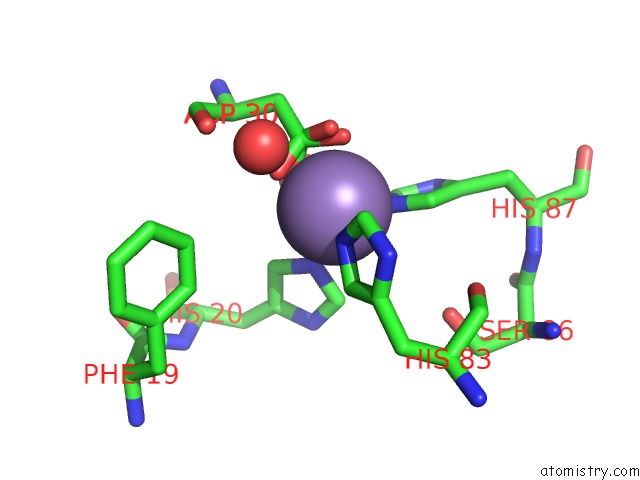
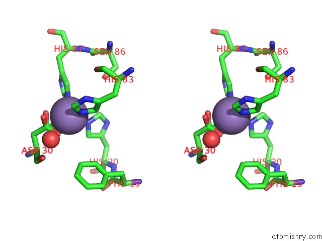
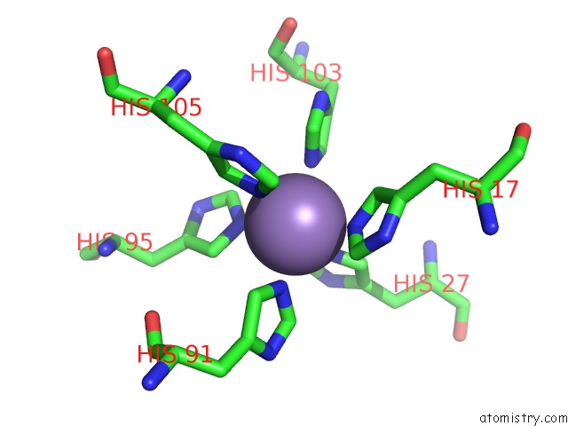
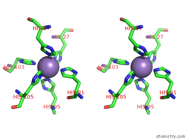
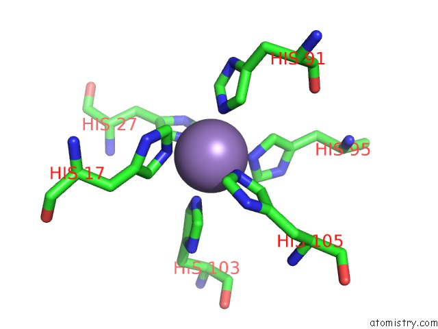
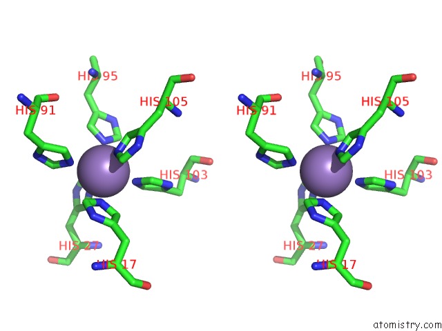
Manganese binding site 1 out of 7 in 4ggf
Go back to
Manganese binding site 1 out
of 7 in the Crystal Structure of MN2+ Bound Calprotectin
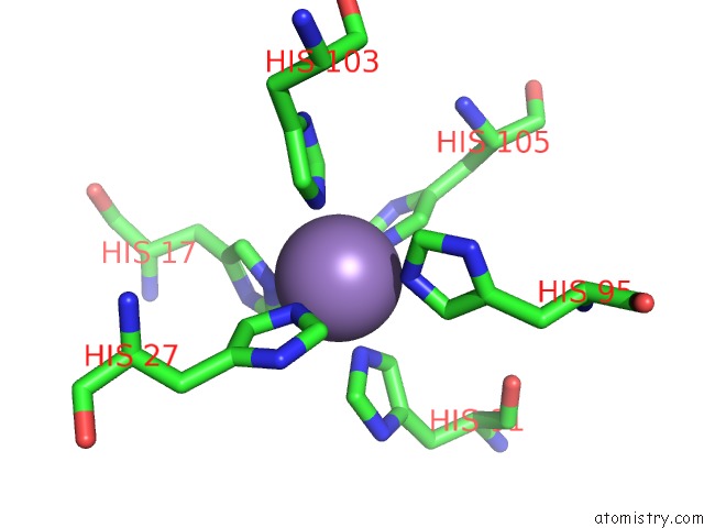
Mono view
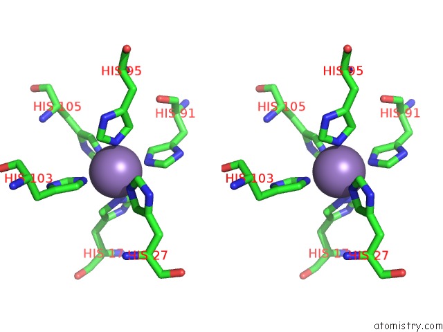
Stereo pair view
Mono view
Stereo pair view
A full contact list of Manganese with other atoms in the Mn binding
site number 1 of Crystal Structure of MN2+ Bound Calprotectin within 5.0Å range:
|
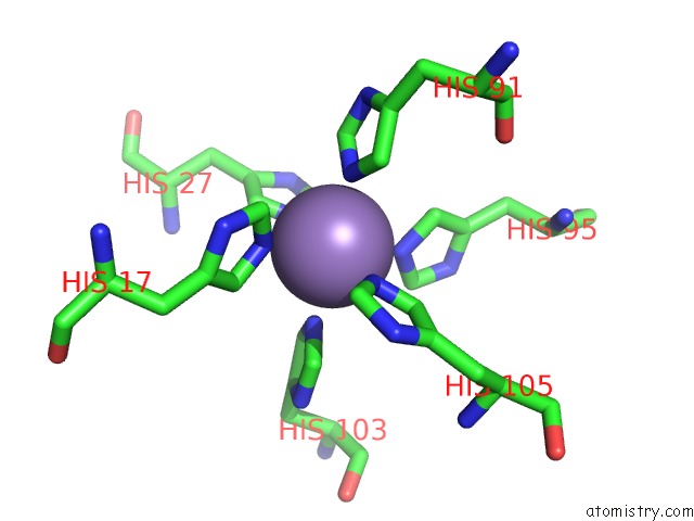
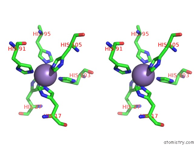
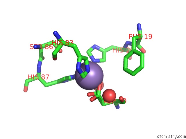
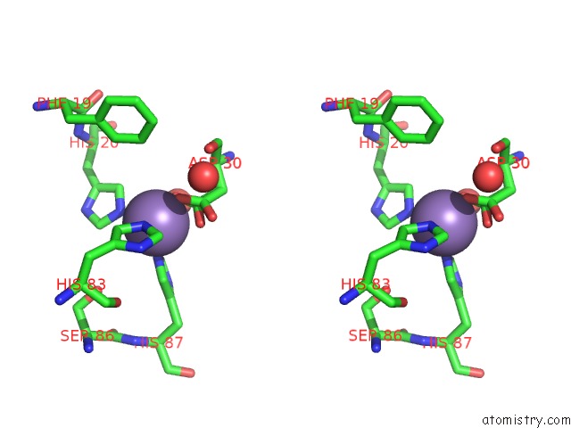
Manganese binding site 2 out of 7 in 4ggf
Go back to
Manganese binding site 2 out
of 7 in the Crystal Structure of MN2+ Bound Calprotectin
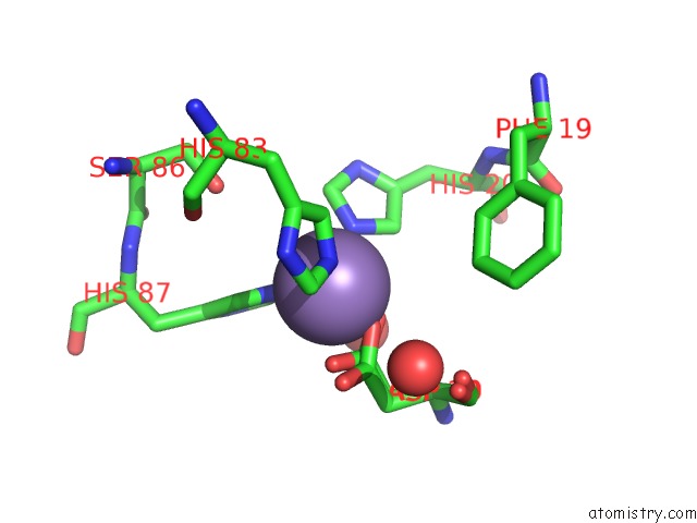
Mono view
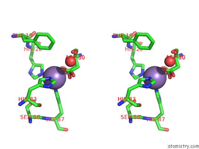
Stereo pair view
Mono view
Stereo pair view
A full contact list of Manganese with other atoms in the Mn binding
site number 2 of Crystal Structure of MN2+ Bound Calprotectin within 5.0Å range:
|
Manganese binding site 3 out of 7 in 4ggf
Go back to
Manganese binding site 3 out
of 7 in the Crystal Structure of MN2+ Bound Calprotectin
Mono view
Stereo pair view
Mono view
Stereo pair view
A full contact list of Manganese with other atoms in the Mn binding
site number 3 of Crystal Structure of MN2+ Bound Calprotectin within 5.0Å range:
|
Manganese binding site 4 out of 7 in 4ggf
Go back to
Manganese binding site 4 out
of 7 in the Crystal Structure of MN2+ Bound Calprotectin
Mono view
Stereo pair view
Mono view
Stereo pair view
A full contact list of Manganese with other atoms in the Mn binding
site number 4 of Crystal Structure of MN2+ Bound Calprotectin within 5.0Å range:
|
Manganese binding site 5 out of 7 in 4ggf
Go back to
Manganese binding site 5 out
of 7 in the Crystal Structure of MN2+ Bound Calprotectin
Mono view
Stereo pair view
Mono view
Stereo pair view
A full contact list of Manganese with other atoms in the Mn binding
site number 5 of Crystal Structure of MN2+ Bound Calprotectin within 5.0Å range:
|
Manganese binding site 6 out of 7 in 4ggf
Go back to
Manganese binding site 6 out
of 7 in the Crystal Structure of MN2+ Bound Calprotectin
Mono view
Stereo pair view
Mono view
Stereo pair view
A full contact list of Manganese with other atoms in the Mn binding
site number 6 of Crystal Structure of MN2+ Bound Calprotectin within 5.0Å range:
|
Manganese binding site 7 out of 7 in 4ggf
Go back to
Manganese binding site 7 out
of 7 in the Crystal Structure of MN2+ Bound Calprotectin
Mono view
Stereo pair view
Mono view
Stereo pair view
A full contact list of Manganese with other atoms in the Mn binding
site number 7 of Crystal Structure of MN2+ Bound Calprotectin within 5.0Å range:
|
Reference:
S.M.Damo,
T.E.Kehl-Fie,
N.Sugitani,
M.E.Holt,
S.Rathi,
W.J.Murphy,
Y.Zhang,
C.Betz,
L.Hench,
G.Fritz,
E.P.Skaar,
W.J.Chazin.
Molecular Basis For Manganese Sequestration By Calprotectin and Roles in the Innate Immune Response to Invading Bacterial Pathogens. Proc.Natl.Acad.Sci.Usa V. 110 3841 2013.
ISSN: ISSN 0027-8424
PubMed: 23431180
DOI: 10.1073/PNAS.1220341110
Page generated: Sat Oct 5 19:30:09 2024
ISSN: ISSN 0027-8424
PubMed: 23431180
DOI: 10.1073/PNAS.1220341110
Last articles
Ca in 2W67Ca in 2W68
Ca in 2W4Z
Ca in 2W4Y
Ca in 2W66
Ca in 2W4X
Ca in 2W3O
Ca in 2W47
Ca in 2W3S
Ca in 2W3R