Manganese »
PDB 4ee3-4fo6 »
4fiy »
Manganese in PDB 4fiy: Crystal Structure of GLFT2 Complexed with Udp
Protein crystallography data
The structure of Crystal Structure of GLFT2 Complexed with Udp, PDB code: 4fiy
was solved by
R.W.Wheatley,
R.B.Zheng,
T.L.Lowary,
K.K.S.Ng,
with X-Ray Crystallography technique. A brief refinement statistics is given in the table below:
| Resolution Low / High (Å) | 40.00 / 3.10 |
| Space group | P 4 21 2 |
| Cell size a, b, c (Å), α, β, γ (°) | 150.398, 150.398, 147.378, 90.00, 90.00, 90.00 |
| R / Rfree (%) | 20.7 / 28.7 |
Manganese Binding Sites:
The binding sites of Manganese atom in the Crystal Structure of GLFT2 Complexed with Udp
(pdb code 4fiy). This binding sites where shown within
5.0 Angstroms radius around Manganese atom.
In total 2 binding sites of Manganese where determined in the Crystal Structure of GLFT2 Complexed with Udp, PDB code: 4fiy:
Jump to Manganese binding site number: 1; 2;
In total 2 binding sites of Manganese where determined in the Crystal Structure of GLFT2 Complexed with Udp, PDB code: 4fiy:
Jump to Manganese binding site number: 1; 2;
Manganese binding site 1 out of 2 in 4fiy
Go back to
Manganese binding site 1 out
of 2 in the Crystal Structure of GLFT2 Complexed with Udp
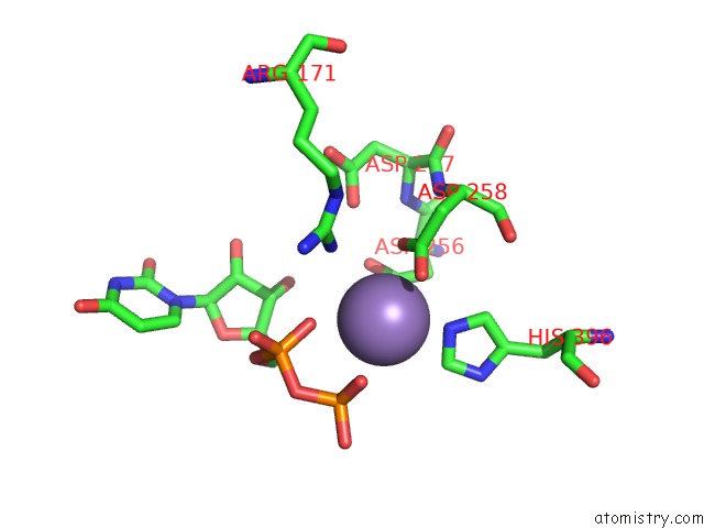
Mono view
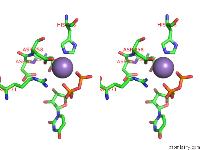
Stereo pair view
Mono view
Stereo pair view
A full contact list of Manganese with other atoms in the Mn binding
site number 1 of Crystal Structure of GLFT2 Complexed with Udp within 5.0Å range:
|
Manganese binding site 2 out of 2 in 4fiy
Go back to
Manganese binding site 2 out
of 2 in the Crystal Structure of GLFT2 Complexed with Udp
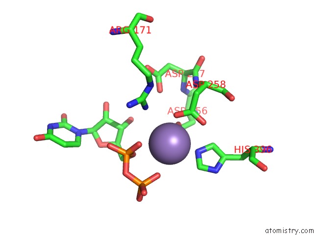
Mono view
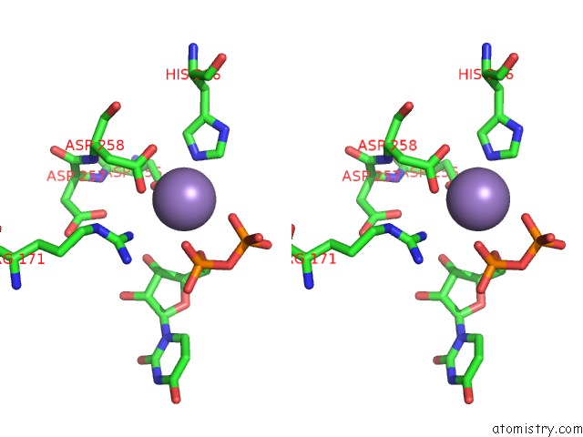
Stereo pair view
Mono view
Stereo pair view
A full contact list of Manganese with other atoms in the Mn binding
site number 2 of Crystal Structure of GLFT2 Complexed with Udp within 5.0Å range:
|
Reference:
R.W.Wheatley,
R.B.Zheng,
M.R.Richards,
T.L.Lowary,
K.K.Ng.
Tetrameric Structure of the GLFT2 Galactofuranosyltransferase Reveals A Scaffold For the Assembly of Mycobacterial Arabinogalactan. J.Biol.Chem. V. 287 28132 2012.
ISSN: ISSN 0021-9258
PubMed: 22707726
DOI: 10.1074/JBC.M112.347484
Page generated: Sat Oct 5 19:25:48 2024
ISSN: ISSN 0021-9258
PubMed: 22707726
DOI: 10.1074/JBC.M112.347484
Last articles
Zn in 9MJ5Zn in 9HNW
Zn in 9G0L
Zn in 9FNE
Zn in 9DZN
Zn in 9E0I
Zn in 9D32
Zn in 9DAK
Zn in 8ZXC
Zn in 8ZUF