Manganese »
PDB 4ee3-4fo6 »
4ewt »
Manganese in PDB 4ewt: The Crystal Structure of A Putative Aminohydrolase From Methicillin Resistant Staphylococcus Aureus
Enzymatic activity of The Crystal Structure of A Putative Aminohydrolase From Methicillin Resistant Staphylococcus Aureus
All present enzymatic activity of The Crystal Structure of A Putative Aminohydrolase From Methicillin Resistant Staphylococcus Aureus:
3.5.1.14;
3.5.1.14;
Protein crystallography data
The structure of The Crystal Structure of A Putative Aminohydrolase From Methicillin Resistant Staphylococcus Aureus, PDB code: 4ewt
was solved by
T.S.Girish,
B.Vivek,
M.Colaco,
S.Misquith,
B.Gopal,
with X-Ray Crystallography technique. A brief refinement statistics is given in the table below:
| Resolution Low / High (Å) | 39.32 / 2.10 |
| Space group | P 1 |
| Cell size a, b, c (Å), α, β, γ (°) | 44.620, 120.110, 132.410, 115.40, 94.64, 96.55 |
| R / Rfree (%) | 19.9 / 22.9 |
Manganese Binding Sites:
The binding sites of Manganese atom in the The Crystal Structure of A Putative Aminohydrolase From Methicillin Resistant Staphylococcus Aureus
(pdb code 4ewt). This binding sites where shown within
5.0 Angstroms radius around Manganese atom.
In total 8 binding sites of Manganese where determined in the The Crystal Structure of A Putative Aminohydrolase From Methicillin Resistant Staphylococcus Aureus, PDB code: 4ewt:
Jump to Manganese binding site number: 1; 2; 3; 4; 5; 6; 7; 8;
In total 8 binding sites of Manganese where determined in the The Crystal Structure of A Putative Aminohydrolase From Methicillin Resistant Staphylococcus Aureus, PDB code: 4ewt:
Jump to Manganese binding site number: 1; 2; 3; 4; 5; 6; 7; 8;
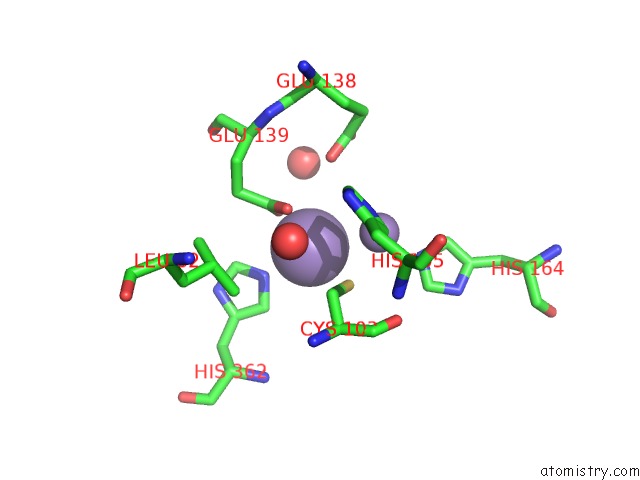
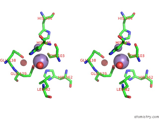
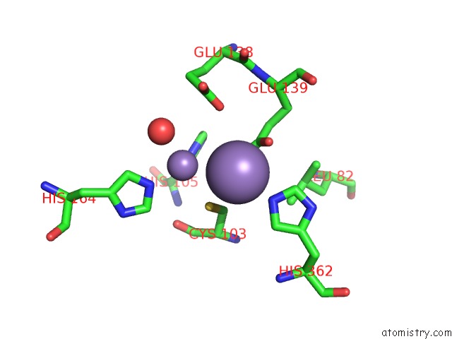
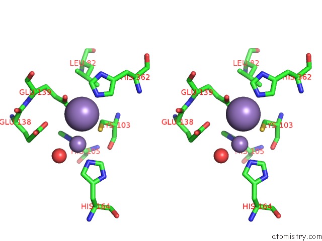
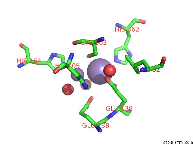
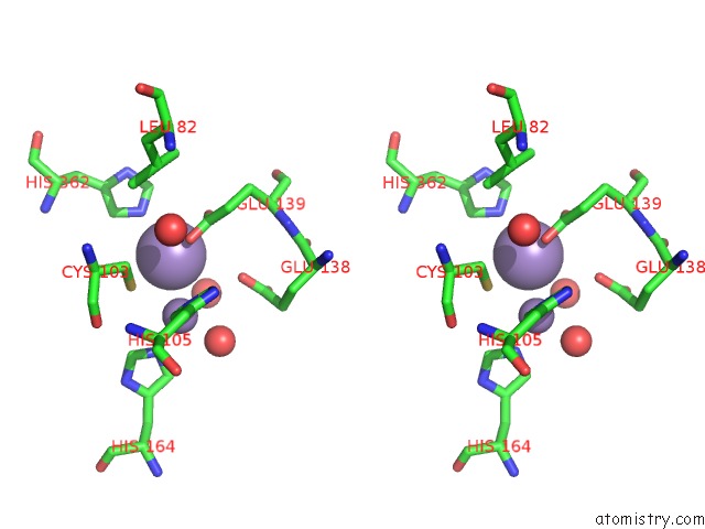
Manganese binding site 1 out of 8 in 4ewt
Go back to
Manganese binding site 1 out
of 8 in the The Crystal Structure of A Putative Aminohydrolase From Methicillin Resistant Staphylococcus Aureus
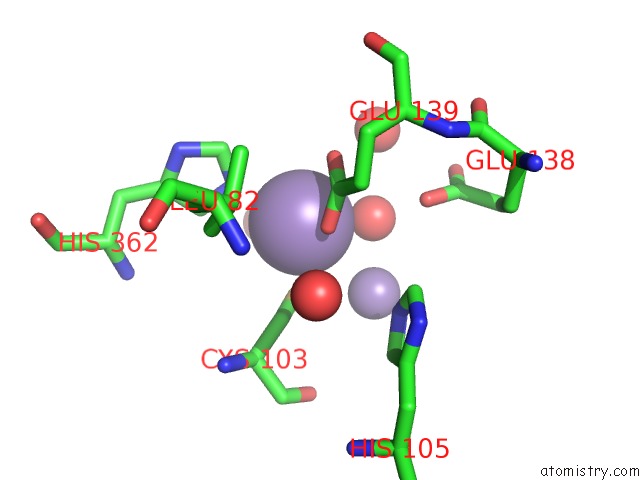
Mono view
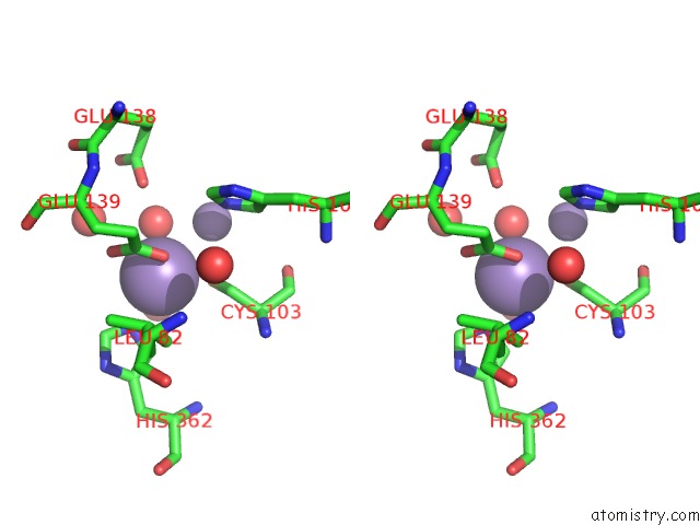
Stereo pair view
Mono view
Stereo pair view
A full contact list of Manganese with other atoms in the Mn binding
site number 1 of The Crystal Structure of A Putative Aminohydrolase From Methicillin Resistant Staphylococcus Aureus within 5.0Å range:
|
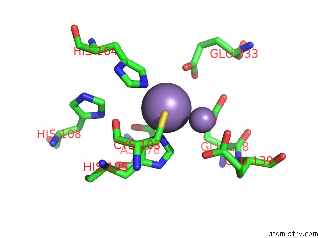
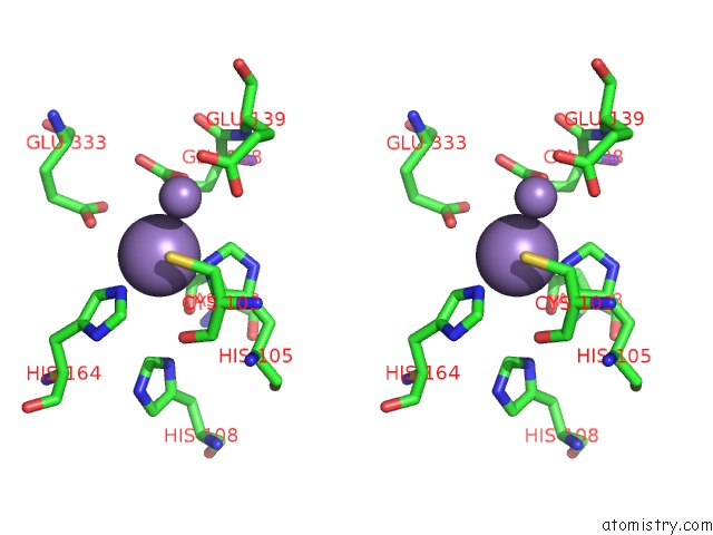
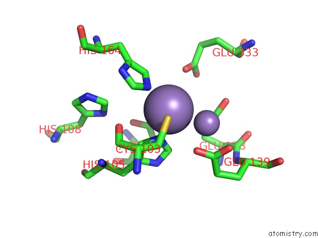
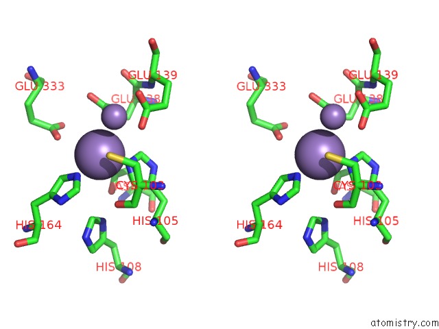
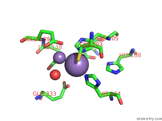
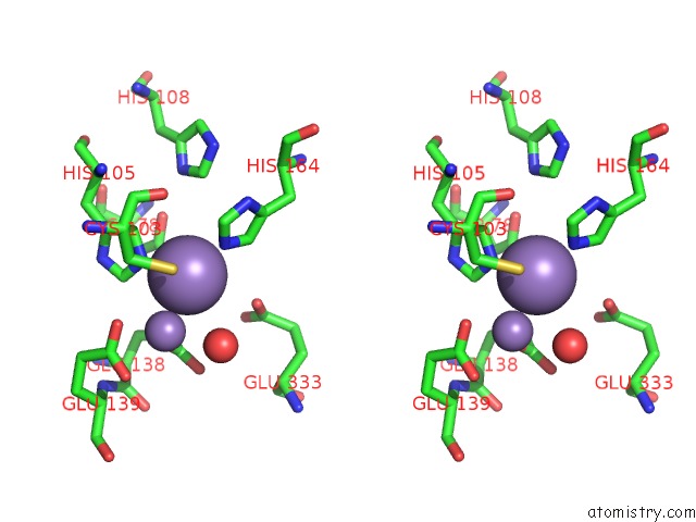
Manganese binding site 2 out of 8 in 4ewt
Go back to
Manganese binding site 2 out
of 8 in the The Crystal Structure of A Putative Aminohydrolase From Methicillin Resistant Staphylococcus Aureus
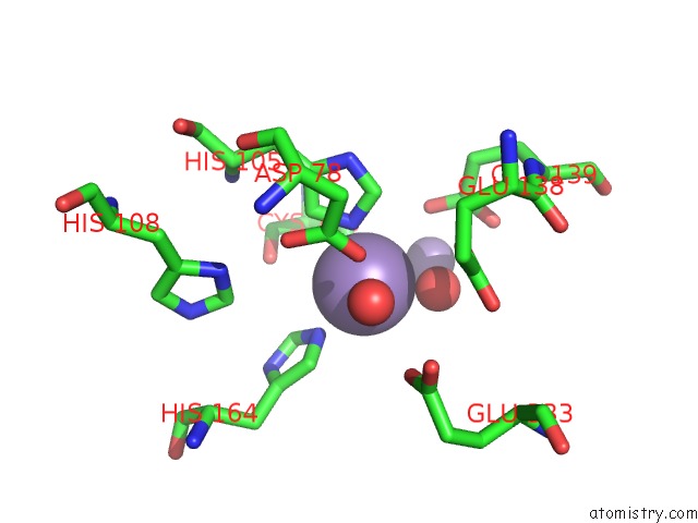
Mono view
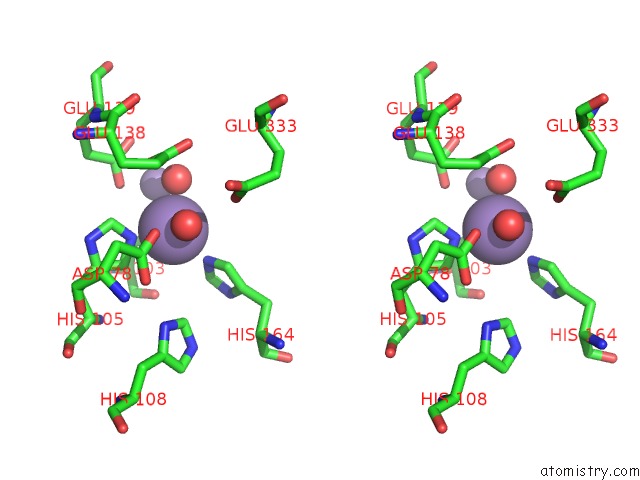
Stereo pair view
Mono view
Stereo pair view
A full contact list of Manganese with other atoms in the Mn binding
site number 2 of The Crystal Structure of A Putative Aminohydrolase From Methicillin Resistant Staphylococcus Aureus within 5.0Å range:
|
Manganese binding site 3 out of 8 in 4ewt
Go back to
Manganese binding site 3 out
of 8 in the The Crystal Structure of A Putative Aminohydrolase From Methicillin Resistant Staphylococcus Aureus
Mono view
Stereo pair view
Mono view
Stereo pair view
A full contact list of Manganese with other atoms in the Mn binding
site number 3 of The Crystal Structure of A Putative Aminohydrolase From Methicillin Resistant Staphylococcus Aureus within 5.0Å range:
|
Manganese binding site 4 out of 8 in 4ewt
Go back to
Manganese binding site 4 out
of 8 in the The Crystal Structure of A Putative Aminohydrolase From Methicillin Resistant Staphylococcus Aureus
Mono view
Stereo pair view
Mono view
Stereo pair view
A full contact list of Manganese with other atoms in the Mn binding
site number 4 of The Crystal Structure of A Putative Aminohydrolase From Methicillin Resistant Staphylococcus Aureus within 5.0Å range:
|
Manganese binding site 5 out of 8 in 4ewt
Go back to
Manganese binding site 5 out
of 8 in the The Crystal Structure of A Putative Aminohydrolase From Methicillin Resistant Staphylococcus Aureus
Mono view
Stereo pair view
Mono view
Stereo pair view
A full contact list of Manganese with other atoms in the Mn binding
site number 5 of The Crystal Structure of A Putative Aminohydrolase From Methicillin Resistant Staphylococcus Aureus within 5.0Å range:
|
Manganese binding site 6 out of 8 in 4ewt
Go back to
Manganese binding site 6 out
of 8 in the The Crystal Structure of A Putative Aminohydrolase From Methicillin Resistant Staphylococcus Aureus
Mono view
Stereo pair view
Mono view
Stereo pair view
A full contact list of Manganese with other atoms in the Mn binding
site number 6 of The Crystal Structure of A Putative Aminohydrolase From Methicillin Resistant Staphylococcus Aureus within 5.0Å range:
|
Manganese binding site 7 out of 8 in 4ewt
Go back to
Manganese binding site 7 out
of 8 in the The Crystal Structure of A Putative Aminohydrolase From Methicillin Resistant Staphylococcus Aureus
Mono view
Stereo pair view
Mono view
Stereo pair view
A full contact list of Manganese with other atoms in the Mn binding
site number 7 of The Crystal Structure of A Putative Aminohydrolase From Methicillin Resistant Staphylococcus Aureus within 5.0Å range:
|
Manganese binding site 8 out of 8 in 4ewt
Go back to
Manganese binding site 8 out
of 8 in the The Crystal Structure of A Putative Aminohydrolase From Methicillin Resistant Staphylococcus Aureus
Mono view
Stereo pair view
Mono view
Stereo pair view
A full contact list of Manganese with other atoms in the Mn binding
site number 8 of The Crystal Structure of A Putative Aminohydrolase From Methicillin Resistant Staphylococcus Aureus within 5.0Å range:
|
Reference:
T.S.Girish,
B.Vivek,
M.Colaco,
S.Misquith,
B.Gopal.
Structure of An Amidohydrolase, SACOL0085, From Methicillin-Resistant Staphylococcus Aureus Col Acta Crystallogr.,Sect.F V. 69 103 2013.
ISSN: ESSN 1744-3091
PubMed: 23385746
DOI: 10.1107/S1744309112049822
Page generated: Sat Oct 5 19:20:52 2024
ISSN: ESSN 1744-3091
PubMed: 23385746
DOI: 10.1107/S1744309112049822
Last articles
Zn in 9MJ5Zn in 9HNW
Zn in 9G0L
Zn in 9FNE
Zn in 9DZN
Zn in 9E0I
Zn in 9D32
Zn in 9DAK
Zn in 8ZXC
Zn in 8ZUF