Manganese »
PDB 4ee3-4fo6 »
4efd »
Manganese in PDB 4efd: Crystal Structure of An M17 Aminopeptidase From Trypanosoma Brucei, TB427TMP.02.4440
Protein crystallography data
The structure of Crystal Structure of An M17 Aminopeptidase From Trypanosoma Brucei, TB427TMP.02.4440, PDB code: 4efd
was solved by
A.K.Wernimont,
K.T.Osman,
P.Loppnau,
C.H.Arrowsmith,
A.M.Edwards,
C.Bountra,
R.Hui,
Y.H.Lin,
Structural Genomics Consortium (Sgc),
with X-Ray Crystallography technique. A brief refinement statistics is given in the table below:
| Resolution Low / High (Å) | 30.00 / 2.45 |
| Space group | P 21 21 21 |
| Cell size a, b, c (Å), α, β, γ (°) | 161.916, 161.740, 176.315, 90.00, 90.00, 90.00 |
| R / Rfree (%) | 22.7 / 25.3 |
Other elements in 4efd:
The structure of Crystal Structure of An M17 Aminopeptidase From Trypanosoma Brucei, TB427TMP.02.4440 also contains other interesting chemical elements:
| Sodium | (Na) | 2 atoms |
Manganese Binding Sites:
The binding sites of Manganese atom in the Crystal Structure of An M17 Aminopeptidase From Trypanosoma Brucei, TB427TMP.02.4440
(pdb code 4efd). This binding sites where shown within
5.0 Angstroms radius around Manganese atom.
In total 8 binding sites of Manganese where determined in the Crystal Structure of An M17 Aminopeptidase From Trypanosoma Brucei, TB427TMP.02.4440, PDB code: 4efd:
Jump to Manganese binding site number: 1; 2; 3; 4; 5; 6; 7; 8;
In total 8 binding sites of Manganese where determined in the Crystal Structure of An M17 Aminopeptidase From Trypanosoma Brucei, TB427TMP.02.4440, PDB code: 4efd:
Jump to Manganese binding site number: 1; 2; 3; 4; 5; 6; 7; 8;
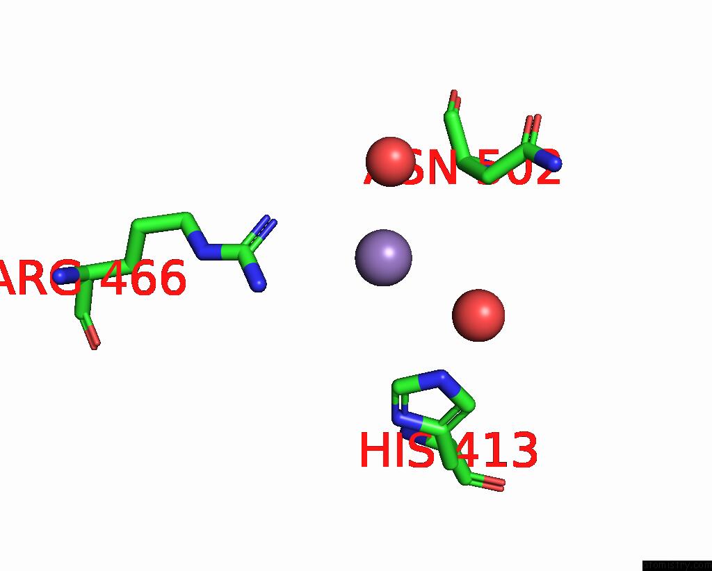
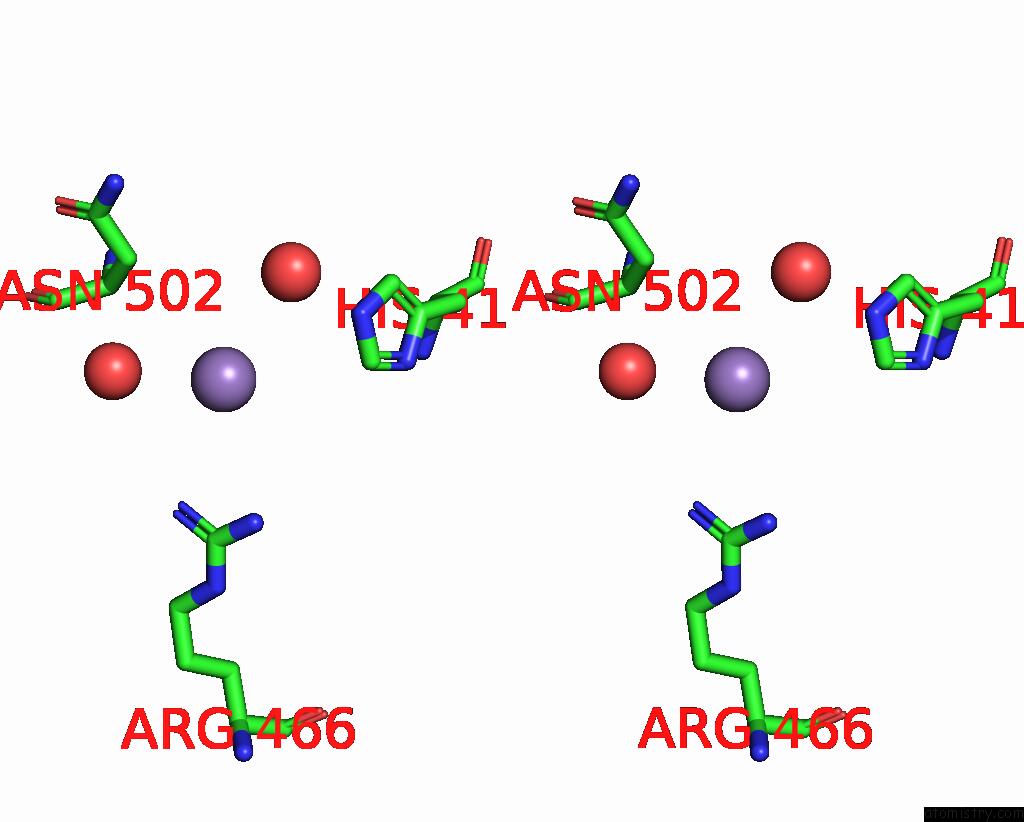
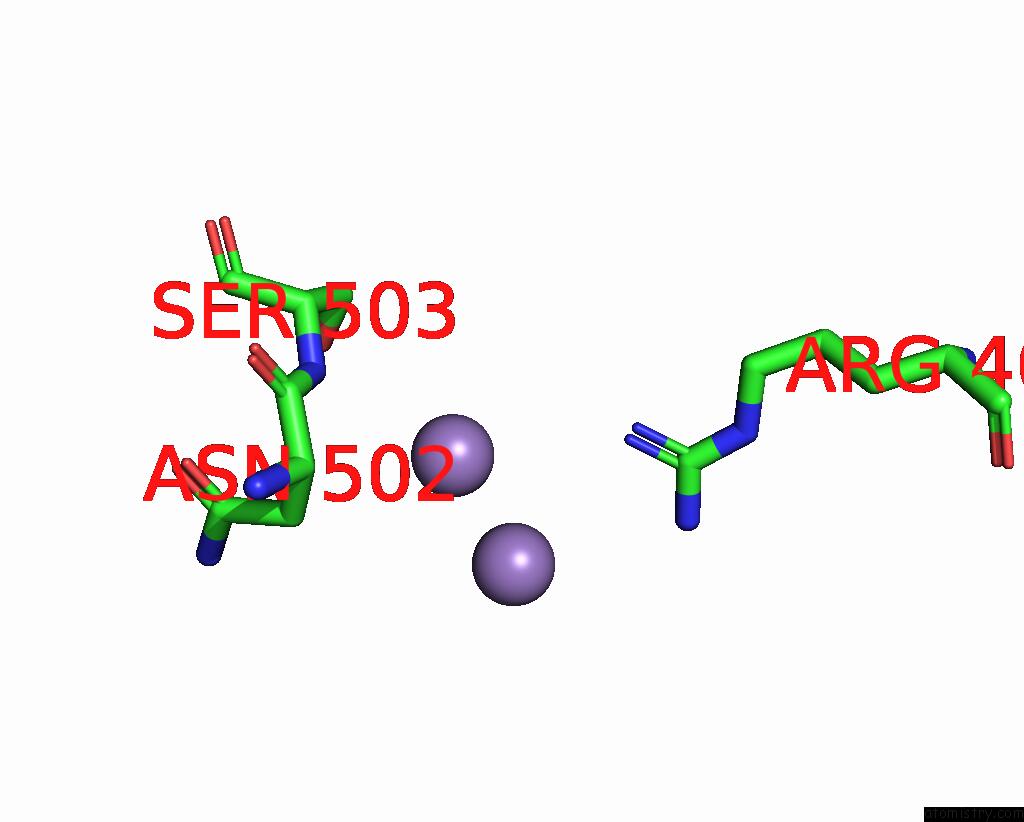
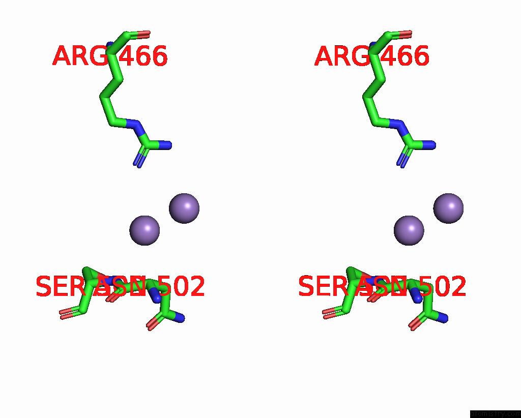
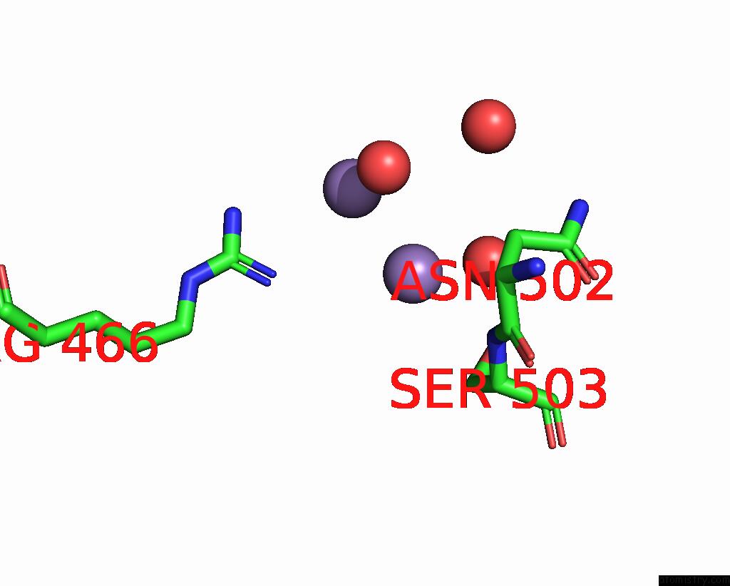
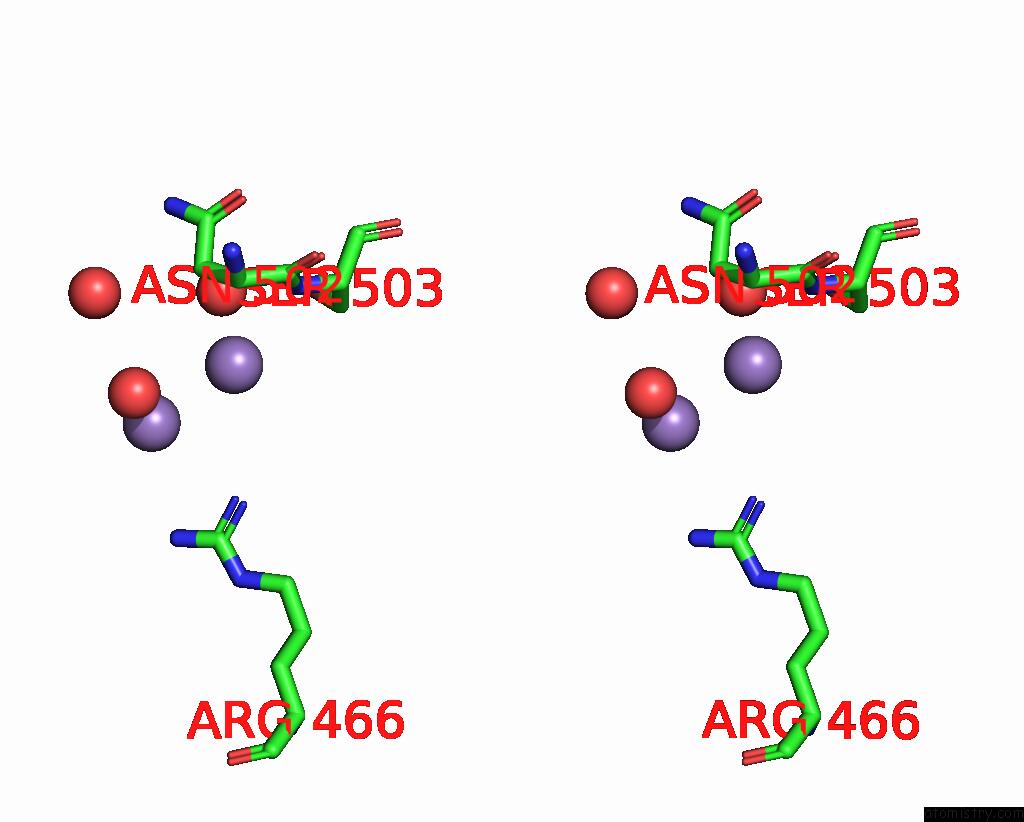
Manganese binding site 1 out of 8 in 4efd
Go back to
Manganese binding site 1 out
of 8 in the Crystal Structure of An M17 Aminopeptidase From Trypanosoma Brucei, TB427TMP.02.4440
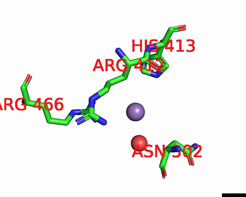
Mono view
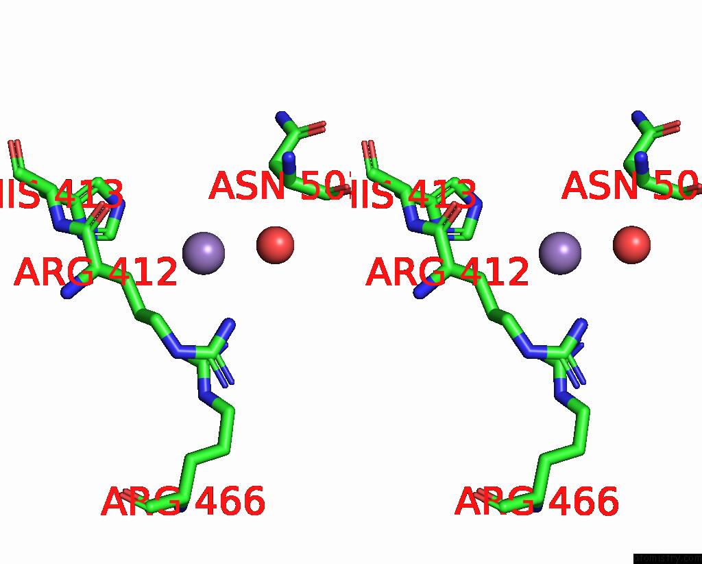
Stereo pair view
Mono view
Stereo pair view
A full contact list of Manganese with other atoms in the Mn binding
site number 1 of Crystal Structure of An M17 Aminopeptidase From Trypanosoma Brucei, TB427TMP.02.4440 within 5.0Å range:
|
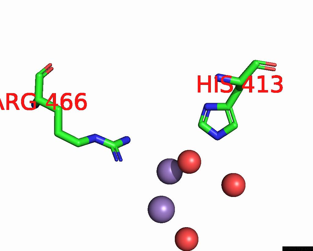
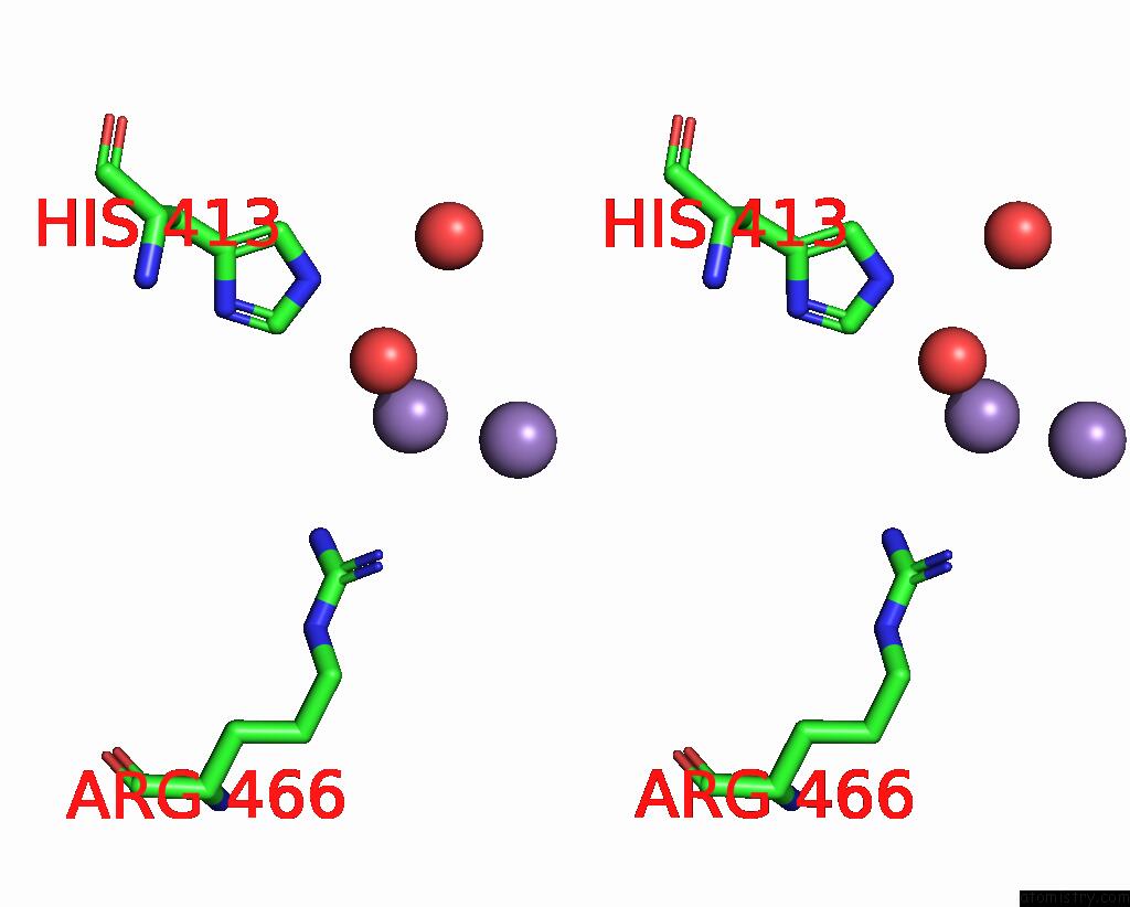
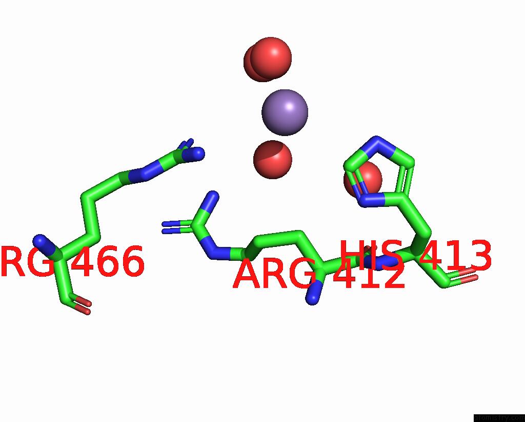
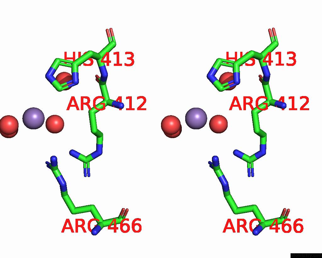
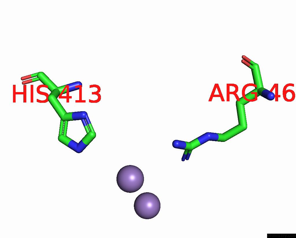
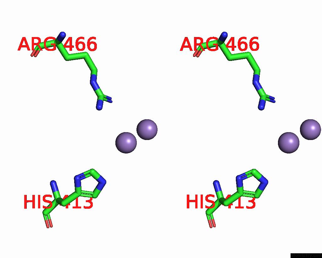
Manganese binding site 2 out of 8 in 4efd
Go back to
Manganese binding site 2 out
of 8 in the Crystal Structure of An M17 Aminopeptidase From Trypanosoma Brucei, TB427TMP.02.4440
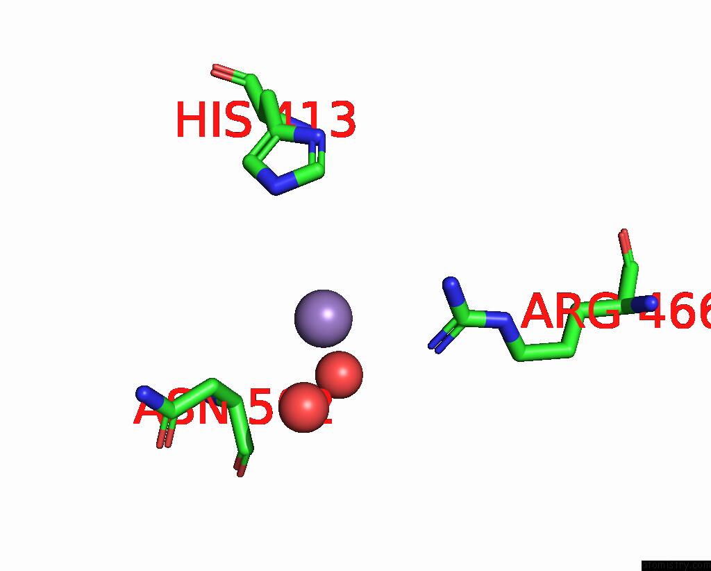
Mono view
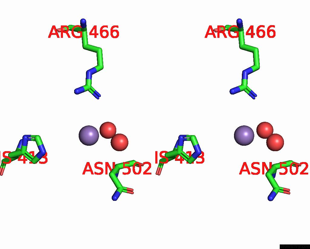
Stereo pair view
Mono view
Stereo pair view
A full contact list of Manganese with other atoms in the Mn binding
site number 2 of Crystal Structure of An M17 Aminopeptidase From Trypanosoma Brucei, TB427TMP.02.4440 within 5.0Å range:
|
Manganese binding site 3 out of 8 in 4efd
Go back to
Manganese binding site 3 out
of 8 in the Crystal Structure of An M17 Aminopeptidase From Trypanosoma Brucei, TB427TMP.02.4440
Mono view
Stereo pair view
Mono view
Stereo pair view
A full contact list of Manganese with other atoms in the Mn binding
site number 3 of Crystal Structure of An M17 Aminopeptidase From Trypanosoma Brucei, TB427TMP.02.4440 within 5.0Å range:
|
Manganese binding site 4 out of 8 in 4efd
Go back to
Manganese binding site 4 out
of 8 in the Crystal Structure of An M17 Aminopeptidase From Trypanosoma Brucei, TB427TMP.02.4440
Mono view
Stereo pair view
Mono view
Stereo pair view
A full contact list of Manganese with other atoms in the Mn binding
site number 4 of Crystal Structure of An M17 Aminopeptidase From Trypanosoma Brucei, TB427TMP.02.4440 within 5.0Å range:
|
Manganese binding site 5 out of 8 in 4efd
Go back to
Manganese binding site 5 out
of 8 in the Crystal Structure of An M17 Aminopeptidase From Trypanosoma Brucei, TB427TMP.02.4440
Mono view
Stereo pair view
Mono view
Stereo pair view
A full contact list of Manganese with other atoms in the Mn binding
site number 5 of Crystal Structure of An M17 Aminopeptidase From Trypanosoma Brucei, TB427TMP.02.4440 within 5.0Å range:
|
Manganese binding site 6 out of 8 in 4efd
Go back to
Manganese binding site 6 out
of 8 in the Crystal Structure of An M17 Aminopeptidase From Trypanosoma Brucei, TB427TMP.02.4440
Mono view
Stereo pair view
Mono view
Stereo pair view
A full contact list of Manganese with other atoms in the Mn binding
site number 6 of Crystal Structure of An M17 Aminopeptidase From Trypanosoma Brucei, TB427TMP.02.4440 within 5.0Å range:
|
Manganese binding site 7 out of 8 in 4efd
Go back to
Manganese binding site 7 out
of 8 in the Crystal Structure of An M17 Aminopeptidase From Trypanosoma Brucei, TB427TMP.02.4440
Mono view
Stereo pair view
Mono view
Stereo pair view
A full contact list of Manganese with other atoms in the Mn binding
site number 7 of Crystal Structure of An M17 Aminopeptidase From Trypanosoma Brucei, TB427TMP.02.4440 within 5.0Å range:
|
Manganese binding site 8 out of 8 in 4efd
Go back to
Manganese binding site 8 out
of 8 in the Crystal Structure of An M17 Aminopeptidase From Trypanosoma Brucei, TB427TMP.02.4440
Mono view
Stereo pair view
Mono view
Stereo pair view
A full contact list of Manganese with other atoms in the Mn binding
site number 8 of Crystal Structure of An M17 Aminopeptidase From Trypanosoma Brucei, TB427TMP.02.4440 within 5.0Å range:
|
Reference:
A.K.Wernimont,
K.T.Osman,
P.Loppnau,
C.H.Arrowsmith,
A.M.Edwards,
C.Bountra,
R.Hui,
Y.H.Lin.
Crystal Structure of An M17 Aminopeptidase From Trypanosoma Brucei, TB427TMP.02.4440 To Be Published.
Page generated: Sat Oct 5 19:18:58 2024
Last articles
Zn in 9MJ5Zn in 9HNW
Zn in 9G0L
Zn in 9FNE
Zn in 9DZN
Zn in 9E0I
Zn in 9D32
Zn in 9DAK
Zn in 8ZXC
Zn in 8ZUF